Copyright
©The Author(s) 2017.
World J Gastroenterol. May 14, 2017; 23(18): 3374-3378
Published online May 14, 2017. doi: 10.3748/wjg.v23.i18.3374
Published online May 14, 2017. doi: 10.3748/wjg.v23.i18.3374
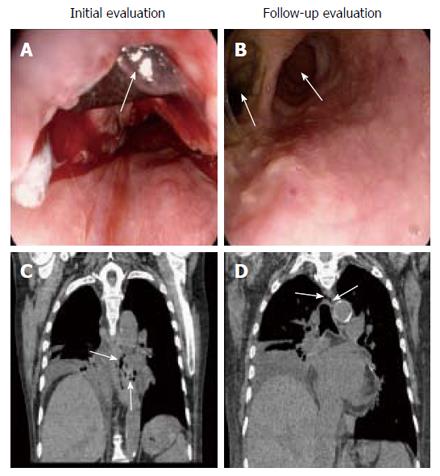
Figure 1 Emergent esophagogastroduodenoscopy.
A: Emergent esophagogastroduodenoscopy (EGD) revealing a large blood clot extending along the esophagus, with an underlying large ulcer (2 cm) in mid esophagus; B: Non-contrast chest CT demonstrating mediastinitis; C: Follow up EGD revealing 4 cm ulceration, with a walled-off false lumen; D: Follow up CT demonstrating false lumen and mediastinitis.
- Citation: Katz-Agranov N, Nevah Rubin MI. Severe esophageal injury after radiofrequency ablation - a deadly complication. World J Gastroenterol 2017; 23(18): 3374-3378
- URL: https://www.wjgnet.com/1007-9327/full/v23/i18/3374.htm
- DOI: https://dx.doi.org/10.3748/wjg.v23.i18.3374


