
















©The Author(s) 2017.
World J Gastroenterol. May 7, 2017; 23(17): 3111-3121
Published online May 7, 2017. doi: 10.3748/wjg.v23.i17.3111
Published online May 7, 2017. doi: 10.3748/wjg.v23.i17.3111
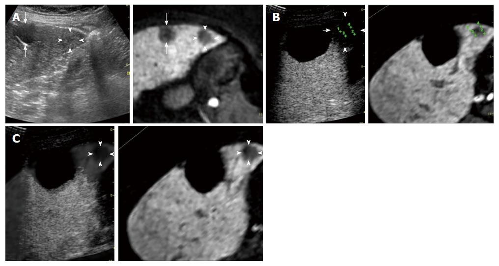
Figure 1 A 70-year-old woman with an early hepatocellular carcinoma lesion (maximum diameter, 18 mm) in segment V of the liver.
A: A fusion image combining conventional ultrasonography (US) (left side) and the hepatobiliary phase of contrast-enhanced magnetic resonance imaging (MRI) with Gd-EOB-DTPA (right side) shows the targeted early hepatocellular carcinoma (HCC) lesion (arrowheads), which appears as an unclear slightly hyper-echoic lesion on conventional US and as a hypo-intense area on the hepatobiliary phase. The arrows indicate the margin of the liver cyst; B: Transcostal fusion imaging, obtained 1 d after radiofrequency ablation (RFA), combining the portal phase of contrast-enhanced US (left side) and the hepatobiliary phase of contrast-enhanced MRI with Gd-EOB-DTPA obtained before RFA (right side) with GPS marks on a single screen. These GPS marks appeared within the ablated areas (arrow), appearing as unenhanced areas on the contrast-enhanced US images. The ablation of the lesion was evaluated as being adequate because of the readily apparent ablative margin; C: Transcostal fusion imaging, obtained 1 d after RFA, combining an overlay of a hepatobiliary phase of contrast-enhanced MRI with Gd-EOB-DTPA on a portal phase of contrast-enhanced US image (left side) and a hepatobiliary phase of contrast-enhanced MRI with Gd-EOB-DTPA obtained before RFA (right side). The edge between the ablated HCC and the ablated adjacent liver parenchyma (arrowheads) is easy to identify with an overlay image.
- Citation: Hao Y, Numata K, Ishii T, Fukuda H, Maeda S, Nakano M, Tanaka K. Rate of local tumor progression following radiofrequency ablation of pathologically early hepatocellular carcinoma. World J Gastroenterol 2017; 23(17): 3111-3121
- URL: https://www.wjgnet.com/1007-9327/full/v23/i17/3111.htm
- DOI: https://dx.doi.org/10.3748/wjg.v23.i17.3111