Copyright
©The Author(s) 2017.
World J Gastroenterol. Apr 28, 2017; 23(16): 3003-3010
Published online Apr 28, 2017. doi: 10.3748/wjg.v23.i16.3003
Published online Apr 28, 2017. doi: 10.3748/wjg.v23.i16.3003
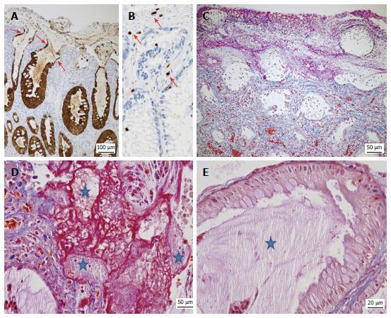
Figure 4 Typical features of protein-losing colitis demonstrated by immunohistochemistry and special stains.
A: Strong cytokeratin 20 expression (brown) by hyperplastic and attenuated (arrows) epithelial cells; B: Detection of rare CD3+ T-cells (brown, arrows) in the lamina propria and epithelium; C: Acid fuchsin orange G-stain (AFOG) stain demonstrating proteins (red) in the apical part of the denuded crypts; the granulation tissue and the pseudomembranes covering the surface, but not in the deeper parts of the mucosa; D, E: AFOG stain combined with a PAS reaction highlighting the presence of both proteins (red) and admixed mucus (pale pink, stars); E: Abundant pale pink mucus (star) but no significant proteins in the crypts lined by hyperplastic goblet cells.
- Citation: Kreisel W, Ruf G, Salm R, Lazaro A, Bengsch B, Globig AM, Fisch P, Lassmann S, Schmitt-Graeff A. Protein-losing pseudomembranous colitis with cap polyposis-like features. World J Gastroenterol 2017; 23(16): 3003-3010
- URL: https://www.wjgnet.com/1007-9327/full/v23/i16/3003.htm
- DOI: https://dx.doi.org/10.3748/wjg.v23.i16.3003


