Copyright
©The Author(s) 2017.
World J Gastroenterol. Apr 28, 2017; 23(16): 3003-3010
Published online Apr 28, 2017. doi: 10.3748/wjg.v23.i16.3003
Published online Apr 28, 2017. doi: 10.3748/wjg.v23.i16.3003
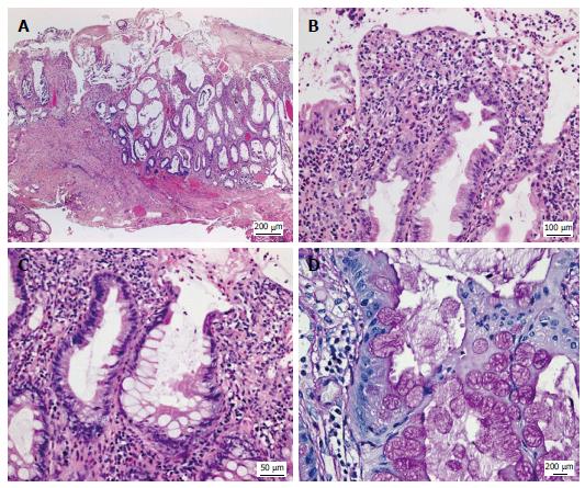
Figure 3 Histologic appearance of the colonic mucosa.
A: Erosive lesions covered by a thick pseudomembranous layer; damage of the surface epithelium and architectural disarray of the mucosa showing prominent cystic dilatation of crypts; B: Foci of mildly serrated epithelium; C, D: Abundant hyperplastic goblet cells associated with a strikingly increased accumulation of intracellular and extracellular mucus. A-C: Hematoxylin and eosin; D: Periodic acid-Schiff reaction.
- Citation: Kreisel W, Ruf G, Salm R, Lazaro A, Bengsch B, Globig AM, Fisch P, Lassmann S, Schmitt-Graeff A. Protein-losing pseudomembranous colitis with cap polyposis-like features. World J Gastroenterol 2017; 23(16): 3003-3010
- URL: https://www.wjgnet.com/1007-9327/full/v23/i16/3003.htm
- DOI: https://dx.doi.org/10.3748/wjg.v23.i16.3003


