Copyright
©The Author(s) 2017.
World J Gastroenterol. Apr 7, 2017; 23(13): 2294-2307
Published online Apr 7, 2017. doi: 10.3748/wjg.v23.i13.2294
Published online Apr 7, 2017. doi: 10.3748/wjg.v23.i13.2294
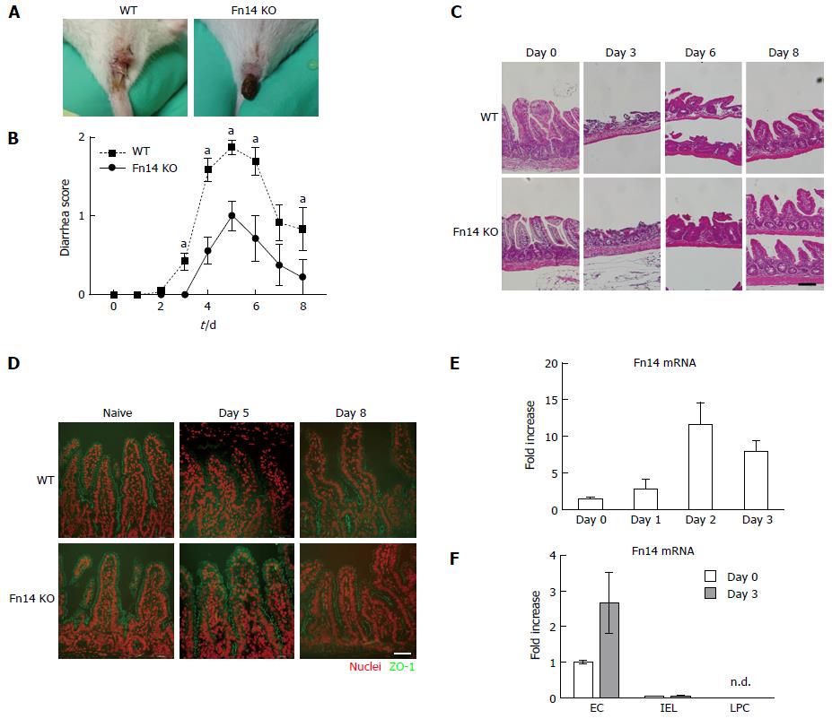
Figure 1 Fn14 deficiency ameliorated 5-FU-induced diarrhea and ileal damage.
A: Representative images of the macroscopic appearance of an anus from a WT mouse with liquid diarrhea and an Fn14 KO mouse with normal fecal pellets following 5-FU administration; B: Diarrhea scores after injection of 5-FU (day 0). Number of mice was about 7-44 at each time point. Results of mouse groups of different observation-time points and endpoints were accumulated and summarized (aP < 0.05); C: Formalin-fixed paraffin-embedded ileal sections were prepared from samples collected at the indicated time point from 5-FU-treated WT or Fn14 KO mice and stained with hematoxylin and eosin. Scale bar = 100 µm; D: Frozen ileal sections were prepared from samples collected at the indicated time point from 5-FU-treated WT or Fn14 KO mice (n = 3 mice per group) and probed with anti-ZO-1 antibody. Green: ZO-1, Red: Nuclear staining. Scale bar = 50 µm. Representative images are shown; E: Fold increase of Fn14 mRNA in total ileal mucosa after injection of 5-FU. Data are presented as the mean ± SD; F: Epithelial cells (EC), intraepithelial lymphocytes (IEL), and lamina propria cells (LPC) were separated from the ileum collected on day 0 (naïve) or day 3 after 5-FU injection, and Fn14 mRNA expression was measured (n = 3 mice per group). Data are presented as the mean ± SD.
- Citation: Sezaki T, Hirata Y, Hagiwara T, Kawamura YI, Okamura T, Takanashi R, Nakano K, Tamura-Nakano M, Burkly LC, Dohi T. Disruption of the TWEAK/Fn14 pathway prevents 5-fluorouracil-induced diarrhea in mice. World J Gastroenterol 2017; 23(13): 2294-2307
- URL: https://www.wjgnet.com/1007-9327/full/v23/i13/2294.htm
- DOI: https://dx.doi.org/10.3748/wjg.v23.i13.2294


