Copyright
©The Author(s) 2017.
World J Gastroenterol. Mar 28, 2017; 23(12): 2106-2123
Published online Mar 28, 2017. doi: 10.3748/wjg.v23.i12.2106
Published online Mar 28, 2017. doi: 10.3748/wjg.v23.i12.2106
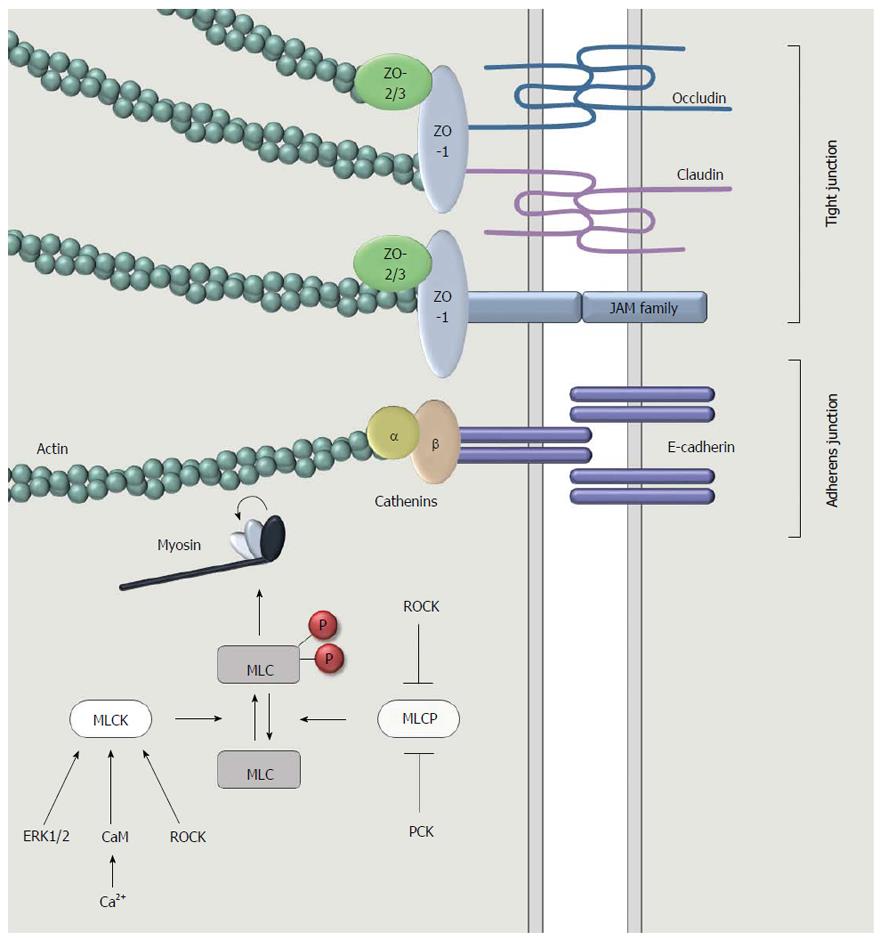
Figure 2 A more detailed representation of the apical junction complex at the intercellular surface between adjacent intestinal epithelial cells.
Tight junctions are comprised of three types of transmembrane proteins: occludin, claudins and junctional adhesion molecules (JAMs). Adaptor proteins such as zonula occludens 1 (ZO-1), ZO-2 and ZO-3 connect the transmembrane proteins to filamentous actin. This cytoskeleton component interacts with myosin to induce a contraction, followed by the opening of the intercellular space. Myosin light chain (MLC) is the main regulator of this contractile machinery. Contraction occurs when MLC is phosphorylated. This is regulated through the activity of myosin light chain kinase (MLCK) and myosin light chain phosphatase (MLCP), which is on their turn regulated by intracellular signaling pathways involving for instance the extracellular signal-regulated kinases (ERK1/2), calcium, calmodulin, protein kinase C (PKC) or Rho-associated protein kinase (ROCK).
- Citation: Van Spaendonk H, Ceuleers H, Witters L, Patteet E, Joossens J, Augustyns K, Lambeir AM, De Meester I, De Man JG, De Winter BY. Regulation of intestinal permeability: The role of proteases. World J Gastroenterol 2017; 23(12): 2106-2123
- URL: https://www.wjgnet.com/1007-9327/full/v23/i12/2106.htm
- DOI: https://dx.doi.org/10.3748/wjg.v23.i12.2106


