Copyright
©The Author(s) 2016.
World J Gastroenterol. Feb 28, 2016; 22(8): 2636-2641
Published online Feb 28, 2016. doi: 10.3748/wjg.v22.i8.2636
Published online Feb 28, 2016. doi: 10.3748/wjg.v22.i8.2636
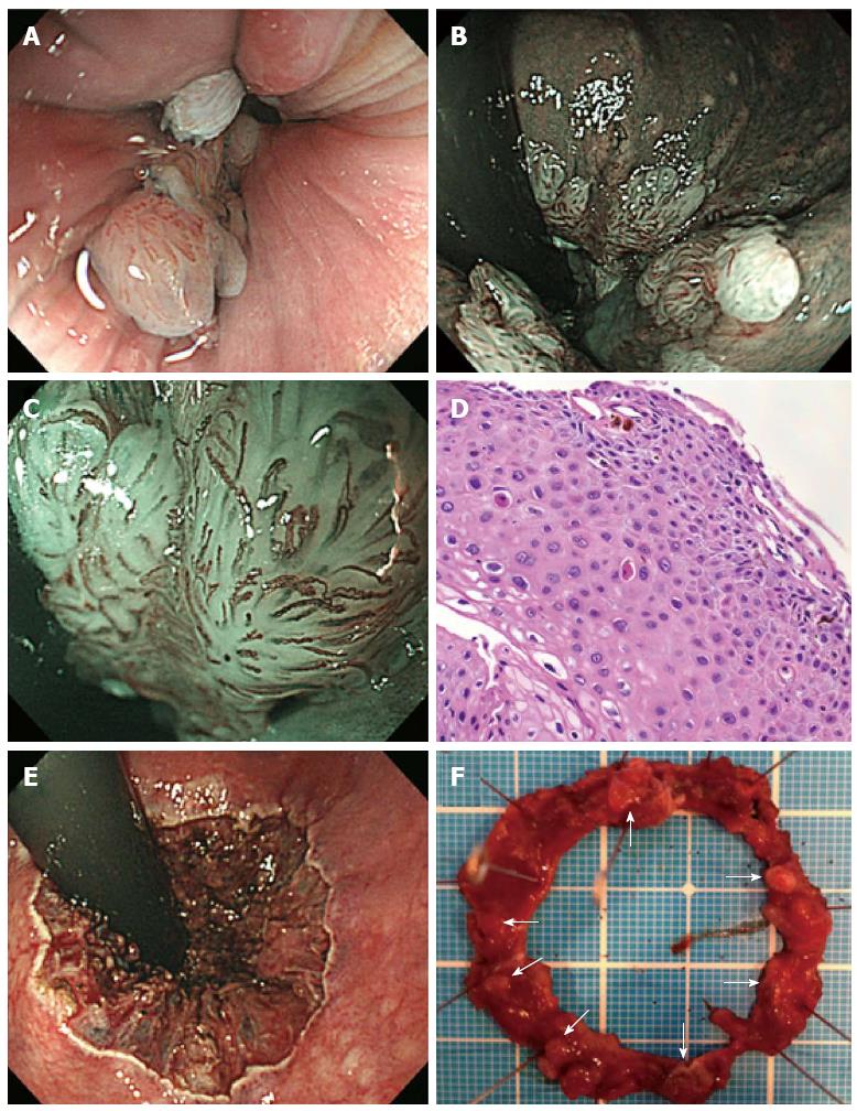
Figure 1 Clinical evaluation of the case of a 28-year-old man who presented with hematochezia.
A: Colonoscopy revealing 10-mm long, whitish condyles extending from the anal canal, with little normal intestinal membrane remaining in the proximal-to-distal site; B: The lesion is almost circumferentially located in the lower rectum, as observed using narrow band imaging (NBI); C: NBI magnification shows dilated red vessels on the surface of the lesion in the anal canal that are indicated as brownish capillaries with hairpin shapes or coil forms; D: Colonic biopsy specimens reveal acanthotic squamous epithelium with hyperpapillomatosis. The epithelial cells show koilocytosis, single cell keratinization, and mild-to-moderate dysplasia of the squamous epithelium. We highly suspected condyloma acuminatum related to human papilloma virus infection; E: Endoscopic submucosal dissection of the lesion all around the anal canal; F: Dissected specimen, 40 mm × 40 mm in diameter. White warts surround almost the entire anal canal (arrow).
- Citation: Sasaki A, Nakajima T, Egashira H, Takeda K, Tokoro S, Ichita C, Masuda S, Uojima H, Koizumi K, Kinbara T, Sakamoto T, Saito Y, Kako M. Condyloma acuminatum of the anal canal, treated with endoscopic submucosal dissection. World J Gastroenterol 2016; 22(8): 2636-2641
- URL: https://www.wjgnet.com/1007-9327/full/v22/i8/2636.htm
- DOI: https://dx.doi.org/10.3748/wjg.v22.i8.2636


