Copyright
©The Author(s) 2016.
World J Gastroenterol. Feb 21, 2016; 22(7): 2398-2402
Published online Feb 21, 2016. doi: 10.3748/wjg.v22.i7.2398
Published online Feb 21, 2016. doi: 10.3748/wjg.v22.i7.2398
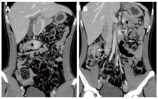
Figure 1 Coronal computed tomography of the abdomen.
A: A cystic lesion 4 cm in diameter in the transverse colon (arrow). The lesion contained stool-like material and acted as a lead point for intussusception; B: Intussusception of the right colon (arrow).
- Citation: Kyo K, Azuma M, Okamoto K, Nishiyama M, Shimamura T, Maema A, Shirakawa M, Nakamura T, Koda K, Yokoyama H. Laparoscopic resection of adult colon duplication causing intussusception. World J Gastroenterol 2016; 22(7): 2398-2402
- URL: https://www.wjgnet.com/1007-9327/full/v22/i7/2398.htm
- DOI: https://dx.doi.org/10.3748/wjg.v22.i7.2398


