Copyright
©The Author(s) 2016.
World J Gastroenterol. Feb 21, 2016; 22(7): 2391-2397
Published online Feb 21, 2016. doi: 10.3748/wjg.v22.i7.2391
Published online Feb 21, 2016. doi: 10.3748/wjg.v22.i7.2391
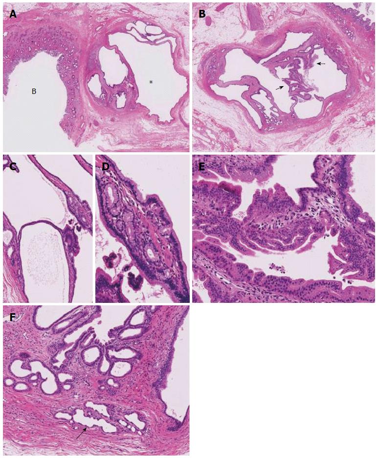
Figure 2 Pathological findings.
A: Low-power histologic image of the same region showing a multilocular cystic lesion (*) and the common hepatic duct involving the carcinoma; B: Another level of a cystic lesion shows a multilocular cystic lesion with some areas exhibiting a micropapillary pattern (arrows), HE; C: The multilocular parts show fine septae lined by columnar epithelium, HE; D: The lining epithelium is columnar with clear cytoplasm, and a pyloric glandular pattern is visible underneath, HE; E: Micropapillary patterns, HE; F: The border of the cystic lesion is rimmed by non-neoplastic peribiliary glands (arrow), HE. HE: Hematoxylin and eosin.
- Citation: Uchida T, Yamamoto Y, Ito T, Okamura Y, Sugiura T, Uesaka K, Nakanuma Y. Cystic micropapillary neoplasm of peribiliary glands with concomitant perihilar cholangiocarcinoma. World J Gastroenterol 2016; 22(7): 2391-2397
- URL: https://www.wjgnet.com/1007-9327/full/v22/i7/2391.htm
- DOI: https://dx.doi.org/10.3748/wjg.v22.i7.2391


