Copyright
©The Author(s) 2016.
World J Gastroenterol. Dec 21, 2016; 22(47): 10287-10303
Published online Dec 21, 2016. doi: 10.3748/wjg.v22.i47.10287
Published online Dec 21, 2016. doi: 10.3748/wjg.v22.i47.10287
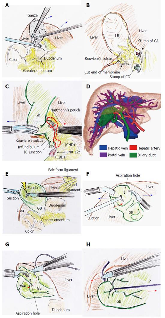
Figure 4 Tips and pitfalls of laparoscopic cholecystectomy.
A: Adequate compression using gauze (blue arrows) works well to stop bleeding at the LB; B: Hemostasis by thermal spread should be never used, nearly at the LB of the GB neck, Rouviere’s sulcus, and CD stump; C: The GB neck and Hartmann’s pouch often extend into the dorsal space due to inflammatory change and/or healing contracture, and unexpected excursions of important ducts and vessels may occur (dotted area). The dissectable/cuttable layer is cut under adequate retraction (blue arrow) as close to the GB as possible using the L-hook electrocautery technique (red arrows); D: Surgeons should not hesitate to perform preoperative detailed imaging studies in complicated cases. The CD (yellow arrow) and CA (red arrow) can be clearly detected on the 3D image; E: The GB is decompressed at the fundus by a dissector with energization; F: Under GB fixation (blue arrows), aspiration is surely performed (red arrow); G: A couple of sutures are placed to close an aspiration hole (dotted arrow); H: The aspiration hole is promptly closed by an extracorporeal ligation (red arrows). CD: Cystic duct; CHD: Common hepatic duct; CVS: Critical view of safety; GB: Gallbladder; IC: Infundibulum-cystic duct; LB: Liver bed; LC: Laparoscopic cholecystectomy.
- Citation: Hori T, Oike F, Furuyama H, Machimoto T, Kadokawa Y, Hata T, Kato S, Yasukawa D, Aisu Y, Sasaki M, Kimura Y, Takamatsu Y, Naito M, Nakauchi M, Tanaka T, Gunji D, Nakamura K, Sato K, Mizuno M, Iida T, Yagi S, Uemoto S, Yoshimura T. Protocol for laparoscopic cholecystectomy: Is it rocket science? World J Gastroenterol 2016; 22(47): 10287-10303
- URL: https://www.wjgnet.com/1007-9327/full/v22/i47/10287.htm
- DOI: https://dx.doi.org/10.3748/wjg.v22.i47.10287


