Copyright
©The Author(s) 2016.
World J Gastroenterol. Dec 21, 2016; 22(47): 10287-10303
Published online Dec 21, 2016. doi: 10.3748/wjg.v22.i47.10287
Published online Dec 21, 2016. doi: 10.3748/wjg.v22.i47.10287
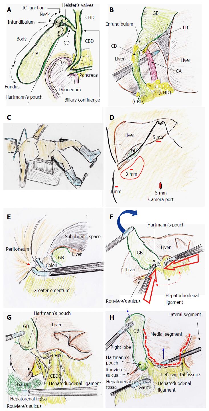
Figure 1 Tips and pitfalls of laparoscopic cholecystectomy.
A: Anatomy is important when performing LC; B: Strasberg’s CVS is shown; C: The patient is placed in a combination of an open-leg supine position and lithotomy position; D: Four ports are placed. The operator’s lateral port should be adequately placed (red circle); E: Port placement (red arrow) should be performed without any injuries; F: The GB fundus is superiorly and cranially lifted (blue arrow). The operator’s ports should then be placed with the forceps tip positioned at an adequate degree around Calot’s triangle (dotted arrow); G: Gauze is placed to dilate the hepatorenal fossa. The hepatoduodenal ligament is stretched. Rouviere’s sulcus and Hartmann’s pouch are confirmed; H: The left sagittal fissure is confirmed. A U-shaped line (dotted line) is visually traced from the round ligament of the liver to the left side of the GB. The bottom plateau of this U-shaped line necessarily involves the CHD and hepatic hilum. Adequate retractions are performed (blue arrows). CD: Cystic duct; CHD: Common hepatic duct; CVS: Critical view of safety; GB: Gallbladder; IC: Infundibulum-cystic duct; LB: Liver bed; LC: Laparoscopic cholecystectomy.
- Citation: Hori T, Oike F, Furuyama H, Machimoto T, Kadokawa Y, Hata T, Kato S, Yasukawa D, Aisu Y, Sasaki M, Kimura Y, Takamatsu Y, Naito M, Nakauchi M, Tanaka T, Gunji D, Nakamura K, Sato K, Mizuno M, Iida T, Yagi S, Uemoto S, Yoshimura T. Protocol for laparoscopic cholecystectomy: Is it rocket science? World J Gastroenterol 2016; 22(47): 10287-10303
- URL: https://www.wjgnet.com/1007-9327/full/v22/i47/10287.htm
- DOI: https://dx.doi.org/10.3748/wjg.v22.i47.10287


