Copyright
©The Author(s) 2016.
World J Gastroenterol. Dec 7, 2016; 22(45): 9880-9897
Published online Dec 7, 2016. doi: 10.3748/wjg.v22.i45.9880
Published online Dec 7, 2016. doi: 10.3748/wjg.v22.i45.9880
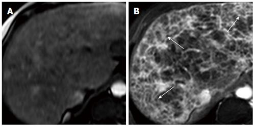
Figure 6 Transverse MR images of cirrhotic liver in vivo[128].
A: SPIO-enhanced two-dimensional spoiled gradient echo (SPGR) image with echotime of 2.65 ms; B: Double-enhanced SPGR image at the same level, showing hyperintense reticulations and hypointense nodules (arrows), thought to represent fibrous septal bands surrounding regenerative nodules.
- Citation: Karanjia RN, Crossey MME, Cox IJ, Fye HKS, Njie R, Goldin RD, Taylor-Robinson SD. Hepatic steatosis and fibrosis: Non-invasive assessment. World J Gastroenterol 2016; 22(45): 9880-9897
- URL: https://www.wjgnet.com/1007-9327/full/v22/i45/9880.htm
- DOI: https://dx.doi.org/10.3748/wjg.v22.i45.9880


