Copyright
©The Author(s) 2016.
World J Gastroenterol. Nov 28, 2016; 22(44): 9860-9864
Published online Nov 28, 2016. doi: 10.3748/wjg.v22.i44.9860
Published online Nov 28, 2016. doi: 10.3748/wjg.v22.i44.9860
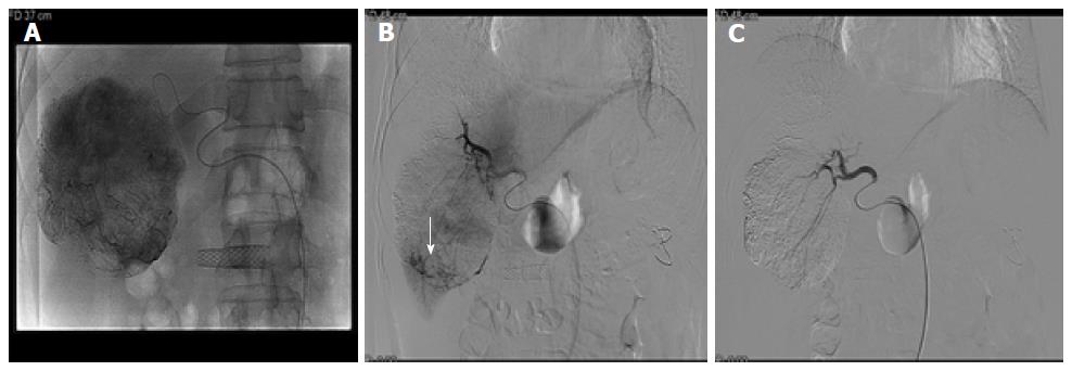
Figure 4 Angiography images.
A: Digital subtraction angiography shows a large ruptured hypervascular tumor with staining at the right hepatic lobe during transarterial embolization; B: Active contrast leakage (white arrow) is seen; C: Selection of the right hepatic artery with a mixture of adriamycin (50 mg) and lipiodol (20 mL). The tumor staining disappears after transarterial embolization.
- Citation: Kim HJ, Park YE, Ki MS, Lee SJ, Beom SH, Han DH, Park YN, Park JY. Spontaneous rupture of hepatic metastasis from a thymoma: A case report. World J Gastroenterol 2016; 22(44): 9860-9864
- URL: https://www.wjgnet.com/1007-9327/full/v22/i44/9860.htm
- DOI: https://dx.doi.org/10.3748/wjg.v22.i44.9860


