Copyright
©The Author(s) 2016.
World J Gastroenterol. Nov 28, 2016; 22(44): 9706-9717
Published online Nov 28, 2016. doi: 10.3748/wjg.v22.i44.9706
Published online Nov 28, 2016. doi: 10.3748/wjg.v22.i44.9706
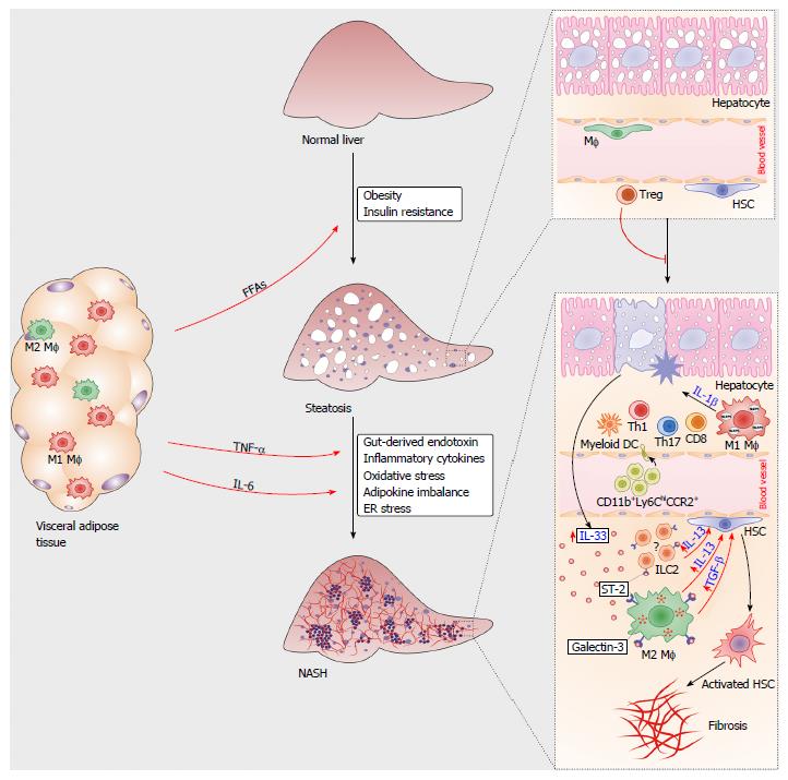
Figure 1 Immune cells in the pathogenesis of nonalcoholic steatohepatitis.
During obesity, proinflammatory M1 macrophages and Th1-type lymphocytes infiltrated in the visceral adipose tissue mediate metaflammation that triggers insulin resistance. Increased amounts of free fatty acids (FFAs) released from adipose tissue accumulate in hepatocytes, causing liver steatosis. Liver regulatory T cells suppress metabolic inflammation. Multiple signals from visceral adipose tissue and gut polarize liver resident macrophages towards M1 type, promote chemotaxis of immune cells and hepatocyte injury. Damaged hepatocytes release IL-33, which promotes release of profibrogenic IL-13 and TGF-β from IL-33R (ST2)-positive macrophages. Liver resident innate lymphoid cells type 2 (ILC2s) might also respond to IL-33 by producing IL-13. Profibrogenic cytokines activate quiescent hepatic stellate cells, which transform to myofibroblasts, the key cells involved in the development of liver fibrosis.
- Citation: Pejnovic N, Jeftic I, Jovicic N, Arsenijevic N, Lukic ML. Galectin-3 and IL-33/ST2 axis roles and interplay in diet-induced steatohepatitis. World J Gastroenterol 2016; 22(44): 9706-9717
- URL: https://www.wjgnet.com/1007-9327/full/v22/i44/9706.htm
- DOI: https://dx.doi.org/10.3748/wjg.v22.i44.9706


