Copyright
©The Author(s) 2016.
World J Gastroenterol. Nov 7, 2016; 22(41): 9229-9234
Published online Nov 7, 2016. doi: 10.3748/wjg.v22.i41.9229
Published online Nov 7, 2016. doi: 10.3748/wjg.v22.i41.9229
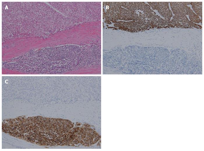
Figure 2 Microscopic findings.
A: Moderately differentiated hepatocellular carcinoma (HCC) is found in the upper portion. The malignant cells show a clear and rich cytoplasm. It is separated from neuroendocrine carcinoma (NEC) by a fibrous band. Poorly differentiated NEC is found in the lower portion. The cytoplasm is barely seen, and the N/C (nucleus/cytoplasm) is very high (hematoxylin eosin staining, magnification × 100); B: Immunohistochemical staining of hepatocyte paraffin-1 is positive in the upper HCC portion (magnification × 100); C: Immunohistochemical staining of CD56 is positive in the lower NEC portion (magnification × 100).
- Citation: Choi GH, Ann SY, Lee SI, Kim SB, Song IH. Collision tumor of hepatocellular carcinoma and neuroendocrine carcinoma involving the liver: Case report and review of the literature. World J Gastroenterol 2016; 22(41): 9229-9234
- URL: https://www.wjgnet.com/1007-9327/full/v22/i41/9229.htm
- DOI: https://dx.doi.org/10.3748/wjg.v22.i41.9229


