Copyright
©The Author(s) 2016.
World J Gastroenterol. Nov 7, 2016; 22(41): 9222-9228
Published online Nov 7, 2016. doi: 10.3748/wjg.v22.i41.9222
Published online Nov 7, 2016. doi: 10.3748/wjg.v22.i41.9222
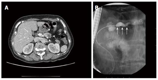
Figure 4 Enhanced abdominal computed tomography scan (A) and endoscopic retrograde cholangiopancreatography image (B) of case 2.
A: The hypovascular tumor in the pancreatic body (arrowheads); B: Dilation and disruption of the main pancreatic duct (arrows).
- Citation: Fujita Y, Kitago M, Masugi Y, Itano O, Shinoda M, Abe Y, Hibi T, Yagi H, Fujii-Nishimura Y, Sakamoto M, Kitagawa Y. Two cases of pancreatic ductal adenocarcinoma with intrapancreatic metastasis. World J Gastroenterol 2016; 22(41): 9222-9228
- URL: https://www.wjgnet.com/1007-9327/full/v22/i41/9222.htm
- DOI: https://dx.doi.org/10.3748/wjg.v22.i41.9222


