Copyright
©The Author(s) 2016.
World J Gastroenterol. Nov 7, 2016; 22(41): 9141-9153
Published online Nov 7, 2016. doi: 10.3748/wjg.v22.i41.9141
Published online Nov 7, 2016. doi: 10.3748/wjg.v22.i41.9141
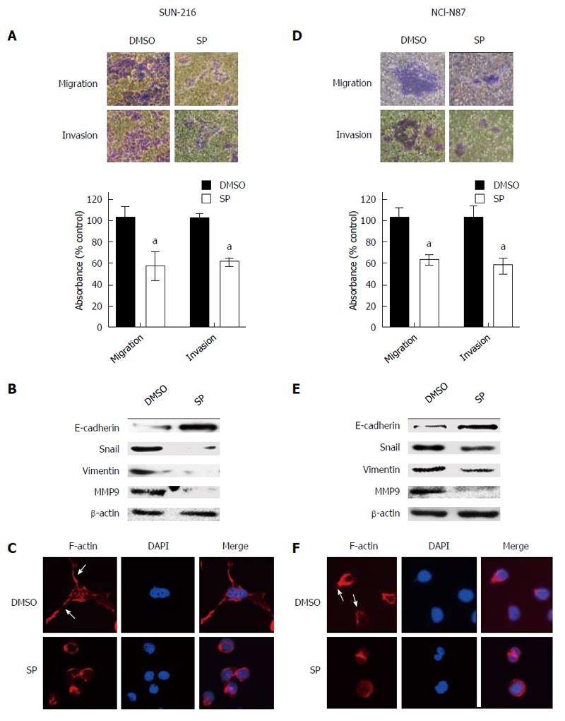
Figure 3 Effect of pharmacological inhibition of JNK on cell migration, invasion, EMT marker expressions and actin cytoskeleton organization.
SNU-216 and NCI-N87 cells were treated with either DMSO or SP600125 (SP). A, D: Cell migration and invasion were analyzed by Transwell assay followed by cell viability assessment using the crystal violet assay. Representative images of migrated/invasive cells taken 48 h after plating into a transwell insert are on the upper, and the quantification of migrated/invasive cells is on the lower. Results were calculated as percentages relative to DMSO vehicle control. Data are expressed as mean ± SD (n = 4 per each group). aP < 0.05 vs DMSO vehicle control; B, E: EMT marker expressions were determined by Western blot; C, F: The organization of the actin cytoskeleton was determined by immunofluorescence staining. Alexa Fluor 633-conjugated phalloidin was used to visualize F-actin (red), and DAPI staining (blue) was used for visualization of cell nuclei. Arrows indicate the FITC-labelled filopodia-like projections. Photographs were taken with a fluorescence microscope. Original magnification, × 400.
- Citation: Choi Y, Ko YS, Park J, Choi Y, Kim Y, Pyo JS, Jang BG, Hwang DH, Kim WH, Lee BL. HER2-induced metastasis is mediated by AKT/JNK/EMT signaling pathway in gastric cancer. World J Gastroenterol 2016; 22(41): 9141-9153
- URL: https://www.wjgnet.com/1007-9327/full/v22/i41/9141.htm
- DOI: https://dx.doi.org/10.3748/wjg.v22.i41.9141


