Copyright
©The Author(s) 2016.
World J Gastroenterol. Nov 7, 2016; 22(41): 9141-9153
Published online Nov 7, 2016. doi: 10.3748/wjg.v22.i41.9141
Published online Nov 7, 2016. doi: 10.3748/wjg.v22.i41.9141
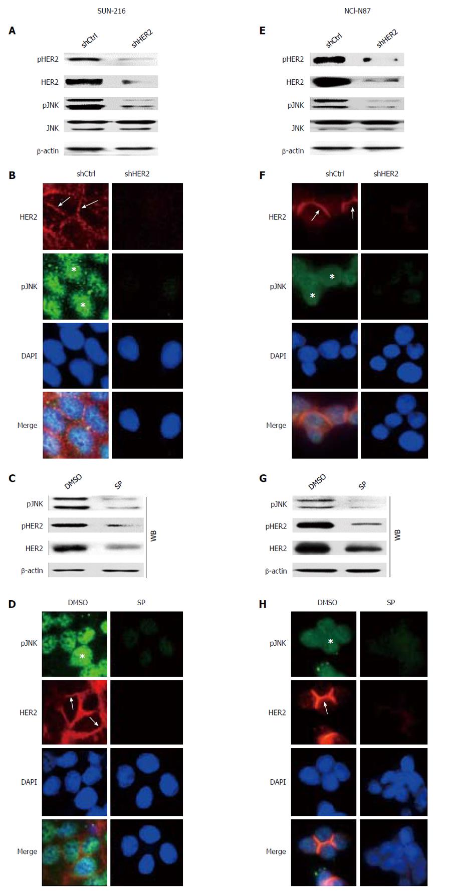
Figure 2 Relationship between HER2 and JNK in SNU-216 and NCI-N87 cells.
A, B, E and F: Cancer cells were infected with a lentivirus containing either control shRNA (shCtrl) or HER2 shRNA (shHER2); A, E: Protein expressions of pHER2, HER2, pJNK and JNK were determined by Western blot; B, F: Double immunofluorescence staining for HER2 (red, arrows) and pJNK (green, asterisks) was performed. Cell nuclei were visualized by DAPI staining (blue). Original magnification, × 400; C, D, G, H: Cells were treated with either DMSO (vehicle control) or SP600125 (SP); C, G: Protein expressions of pJNK, pHER2 and HER2 were determined by Western blot (WB) and HER2 mRNA expression was determined by semi-quantitative reverse transcription-polymerase chain reaction (RT-PCR); D, H: Double immunofluorescence staining for pJNK (green, asterisks) and HER2 (red, arrows) was performed. Cell nuclei were visualized by DAPI staining (blue). Original magnification, × 400.
- Citation: Choi Y, Ko YS, Park J, Choi Y, Kim Y, Pyo JS, Jang BG, Hwang DH, Kim WH, Lee BL. HER2-induced metastasis is mediated by AKT/JNK/EMT signaling pathway in gastric cancer. World J Gastroenterol 2016; 22(41): 9141-9153
- URL: https://www.wjgnet.com/1007-9327/full/v22/i41/9141.htm
- DOI: https://dx.doi.org/10.3748/wjg.v22.i41.9141


