
















©The Author(s) 2016.
World J Gastroenterol. Nov 7, 2016; 22(41): 9127-9140
Published online Nov 7, 2016. doi: 10.3748/wjg.v22.i41.9127
Published online Nov 7, 2016. doi: 10.3748/wjg.v22.i41.9127
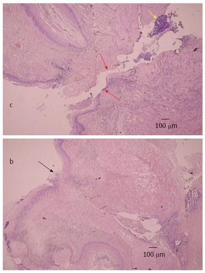
Figure 7 Illustrative microscopic presentation in rats that underwent esophagogastric anastomosis at post-operative day 2, hematoxylin eosin staining.
Separated anastomosis edges (red arrows) with neutrophils within the edges (yellow arrow) were consistently noted in controls (c). Separated wound edges (red arrows). Connected edges in rats that underwent esophagogastric anastomosis and treated with BPC 157 regimens (black arrow) (b).
- Citation: Djakovic Z, Djakovic I, Cesarec V, Madzarac G, Becejac T, Zukanovic G, Drmic D, Batelja L, Zenko Sever A, Kolenc D, Pajtak A, Knez N, Japjec M, Luetic K, Stancic-Rokotov D, Seiwerth S, Sikiric P. Esophagogastric anastomosis in rats: Improved healing by BPC 157 and L-arginine, aggravated by L-NAME. World J Gastroenterol 2016; 22(41): 9127-9140
- URL: https://www.wjgnet.com/1007-9327/full/v22/i41/9127.htm
- DOI: https://dx.doi.org/10.3748/wjg.v22.i41.9127