Copyright
©The Author(s) 2016.
World J Gastroenterol. Oct 28, 2016; 22(40): 8949-8955
Published online Oct 28, 2016. doi: 10.3748/wjg.v22.i40.8949
Published online Oct 28, 2016. doi: 10.3748/wjg.v22.i40.8949
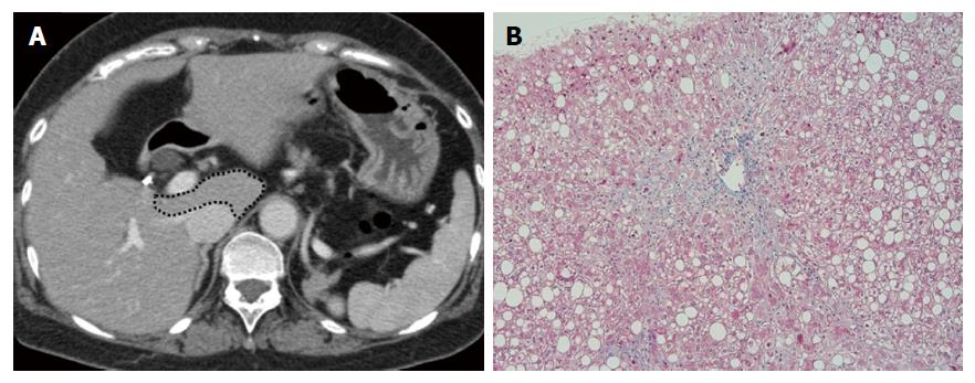
Figure 4 A 63-year-old woman with nonalcoholic steatohepatitis, fibrosis stage 1.
The total volume of the liver was 1190.9 mL, and volume percentages of the left lateral segment, left medial segment, caudate lobe and right lobe were 21.8%, 13.7%, 4.3% and 60.2%, respectively (A). The dotted line shows the caudate lobe. The biopsy specimen showed pericellular fibrosis at zone 3 (B).
- Citation: Fujita N, Nishie A, Asayama Y, Ishigami K, Ushijima Y, Takayama Y, Okamoto D, Shirabe K, Yoshizumi T, Kotoh K, Furusyo N, Hida T, Oda Y, Fujioka T, Honda H. Fibrosis in nonalcoholic fatty liver disease: Noninvasive assessment using computed tomography volumetry. World J Gastroenterol 2016; 22(40): 8949-8955
- URL: https://www.wjgnet.com/1007-9327/full/v22/i40/8949.htm
- DOI: https://dx.doi.org/10.3748/wjg.v22.i40.8949


