Copyright
©The Author(s) 2016.
World J Gastroenterol. Oct 14, 2016; 22(38): 8605-8614
Published online Oct 14, 2016. doi: 10.3748/wjg.v22.i38.8605
Published online Oct 14, 2016. doi: 10.3748/wjg.v22.i38.8605
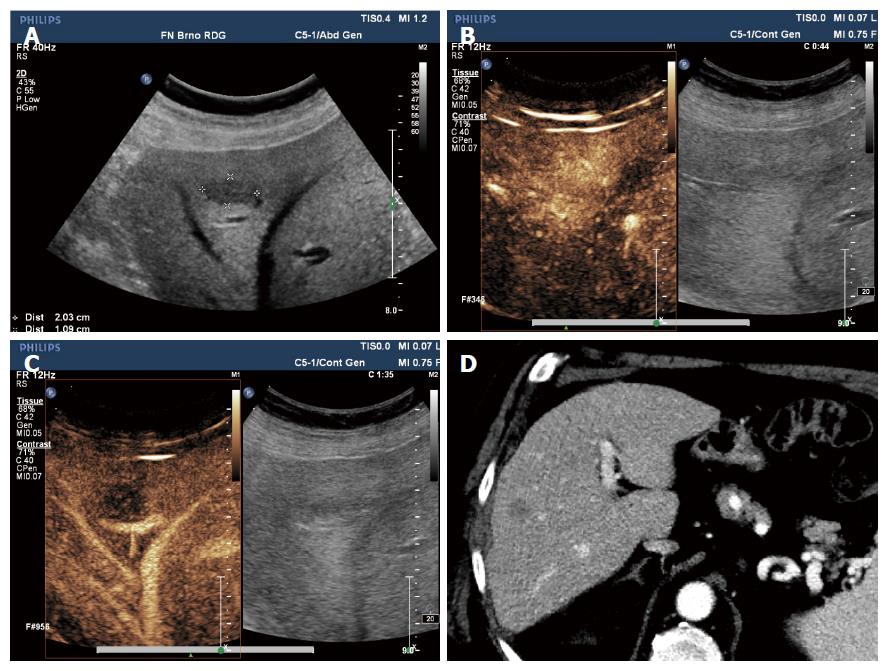
Figure 1 Liver metastasis of spinocellular carcinoma.
Male, 75 years of age, with history of spinocellular carcinoma of the left lung. Emergency ultrasound performed for colicky abdominal pain identifies a hypoechoic lesion in segment S8 of the right liver lobe (A); Upon contrast-enhanced ultrasonography (CEUS) examination, the lesion shows increased enhancement in arterial phase compared to surrounding parenchyma (B); In following phases (C), there is detectable washout typical for malignant lesions. On CT performed for another reason there is a hypodense lesion on portal venous phase (D) corresponding to metastasis.
- Citation: Smajerova M, Petrasova H, Little J, Ovesna P, Andrasina T, Valek V, Nemcova E, Miklosova B. Contrast-enhanced ultrasonography in the evaluation of incidental focal liver lesions: A cost-effectiveness analysis. World J Gastroenterol 2016; 22(38): 8605-8614
- URL: https://www.wjgnet.com/1007-9327/full/v22/i38/8605.htm
- DOI: https://dx.doi.org/10.3748/wjg.v22.i38.8605


