Copyright
©The Author(s) 2016.
World J Gastroenterol. Oct 14, 2016; 22(38): 8596-8604
Published online Oct 14, 2016. doi: 10.3748/wjg.v22.i38.8596
Published online Oct 14, 2016. doi: 10.3748/wjg.v22.i38.8596
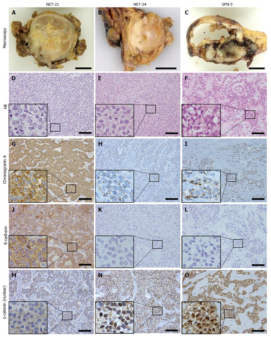
Figure 1 Macroscopic features and representative staining of a typical case of neuroendocrine tumor (case NET-21: A, D, G, J, M), a confusing case (case NET-24: B, E, H, K, N), and a typical case of solid-pseudopapillary neoplasm (case SPN-5: C, F, I, L, O).
The typical NET case showed a solid growth pattern (A) with homogeneous cells, being strongly positive for chromogranin A and E-cadherin, and did not show nuclear labeling of β-catenin. In contrast, the typical SPN case showed a solid and cystic growth pattern (C) containing pseudopapillary structures formed by poorly cohesive cells, with a dot-like pattern of chromogranin A, negativity for E-cadherin, and nuclear labeling for β-catenin. NET-24, originally diagnosed as NET, mimicked the macroscopic features of NET, but exhibited the same immunohistochemical profile as SPN. HE, hematoxylin and eosin stain. Asterisk, cystic lesion of SPN case. Scale bars, 1 cm (macroscopic image) and 100 μm (microscopic image). Small boxes indicate representative magnified fields.
- Citation: Ohara Y, Oda T, Hashimoto S, Akashi Y, Miyamoto R, Enomoto T, Satomi K, Morishita Y, Ohkohchi N. Pancreatic neuroendocrine tumor and solid-pseudopapillary neoplasm: Key immunohistochemical profiles for differential diagnosis. World J Gastroenterol 2016; 22(38): 8596-8604
- URL: https://www.wjgnet.com/1007-9327/full/v22/i38/8596.htm
- DOI: https://dx.doi.org/10.3748/wjg.v22.i38.8596


