Copyright
©The Author(s) 2016.
World J Gastroenterol. Oct 7, 2016; 22(37): 8435-8438
Published online Oct 7, 2016. doi: 10.3748/wjg.v22.i37.8435
Published online Oct 7, 2016. doi: 10.3748/wjg.v22.i37.8435
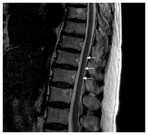
Figure 2 T2-weighted sagittal magnetic resonance image of the thoracic spinal cord in the patient.
The white arrows indicate high signal intensity in the posterior column.
- Citation: Hwang CH, Park DJ, Kim GY. Ataxic gait following total gastrectomy for gastric cancer. World J Gastroenterol 2016; 22(37): 8435-8438
- URL: https://www.wjgnet.com/1007-9327/full/v22/i37/8435.htm
- DOI: https://dx.doi.org/10.3748/wjg.v22.i37.8435


