Copyright
©The Author(s) 2016.
World J Gastroenterol. Oct 7, 2016; 22(37): 8334-8348
Published online Oct 7, 2016. doi: 10.3748/wjg.v22.i37.8334
Published online Oct 7, 2016. doi: 10.3748/wjg.v22.i37.8334
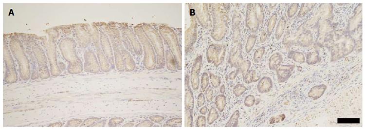
Figure 8 Immunohistochemical detection of Cxcl5 in Winnie distal colonic mucosa.
A: Representative distal colon from Winnie without DSS exposure (n = 6). Cxcl5 labelling displayed a diffuse cytoplasmic pattern in the colonic epithelium; B: Representative distal mucosa of Winnie exposed to DSS (n = 11). Dysplastic glands displaying focal reduction in epithelial Cxcl5. Weak Cxcl5 immunolabelling is present within the cytoplasm of the epithelial cells lining dysplastic glands. Note that submucosal epithelium penetrating the muscularis mucosae retains Cxcl5 expression. Scale bar represents 50 μm.
- Citation: Randall-Demllo S, Fernando R, Brain T, Sohal SS, Cook AL, Guven N, Kunde D, Spring K, Eri R. Characterisation of colonic dysplasia-like epithelial atypia in murine colitis. World J Gastroenterol 2016; 22(37): 8334-8348
- URL: https://www.wjgnet.com/1007-9327/full/v22/i37/8334.htm
- DOI: https://dx.doi.org/10.3748/wjg.v22.i37.8334


