Copyright
©The Author(s) 2016.
World J Gastroenterol. Sep 28, 2016; 22(36): 8242-8246
Published online Sep 28, 2016. doi: 10.3748/wjg.v22.i36.8242
Published online Sep 28, 2016. doi: 10.3748/wjg.v22.i36.8242
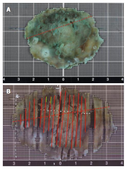
Figure 2 A macroscopic view of the specimen resected by endoscopic submucosal dissection.
A: Macroscopic appearance of the specimen soaked for almost 24 h in formalin after endoscopic submucosal dissection. The red dotted line indicates where there was a pyloric ring; B: Cut out of the resected specimen. The solid red line indicates adenocarcinoma; green line indicates neuroendocrine tumor; red dotted line indicates the location of the pylorus.
- Citation: Kaneko H, Miyake A, Ishii Y, Sue S, Miwa H, Sasaki T, Tamura T, Kondo M, Maeda S. Case of a tumor comprising gastric cancer and duodenal neuroendocrine tumor. World J Gastroenterol 2016; 22(36): 8242-8246
- URL: https://www.wjgnet.com/1007-9327/full/v22/i36/8242.htm
- DOI: https://dx.doi.org/10.3748/wjg.v22.i36.8242


