
















©The Author(s) 2016.
World J Gastroenterol. Sep 14, 2016; 22(34): 7692-7707
Published online Sep 14, 2016. doi: 10.3748/wjg.v22.i34.7692
Published online Sep 14, 2016. doi: 10.3748/wjg.v22.i34.7692
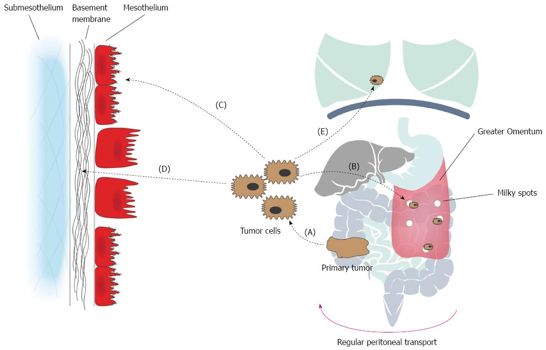
Figure 2 Pathophysiology of colorectal peritoneal carcinomatosis: the peritoneal metastatic cascade.
The emergence of PC is the result of a molecular crosstalk between tumor cells and host elements, comprising several well-defined steps. A: Individual or clumps of tumor cells detach from the primary tumor and gain access to the peritoneal cavity. Spontaneous exfoliation of tumor cells from the primary tumor can be promoted by the down-regulation of E-cadherin, increased interstitial fluid pressure, and iatrogenically during surgery; B: The free tumor cells become susceptible to the regular peritoneal transport. Peritoneal transport is due to changes in the intra-abdominal pressure during respiration, gravity and peristalsis of the bowel; which results in a clockwise flow from the pelvis, along the right paracolic gutter and to the subdiaphragmatic space and finally towards the pelvis again; C: Attachment of tumor cells to distant peritoneum occurs via two processes, denominated transmesothelial and translymphatic metastasis. During transmesothelial metastasis, loose tumor cells directly adhere to distant mesothelium through adhesion molecules. During translymphatic metastasis, free tumor cells gain access to the submesothelial lymphatics through lymphatic stomata. Preferential tumor growth in the milky spots of the greater omentum has been observed; D: Tumor cells invade the submesothelium. In areas of absent or rounded (cuboidal) mesothelial cells, tumor cells interact with the laminar network of the basement membrane through integrin-mediated adhesion. Subsequent invasion of the submesothelial tissue occurs via degradation by proteases (MMPs); E: Systemic metastasis. PC: Peritoneal carcinomatosis.
- Citation: Lemoine L, Sugarbaker P, Van der Speeten K. Pathophysiology of colorectal peritoneal carcinomatosis: Role of the peritoneum. World J Gastroenterol 2016; 22(34): 7692-7707
- URL: https://www.wjgnet.com/1007-9327/full/v22/i34/7692.htm
- DOI: https://dx.doi.org/10.3748/wjg.v22.i34.7692