
















©The Author(s) 2016.
World J Gastroenterol. Sep 14, 2016; 22(34): 7692-7707
Published online Sep 14, 2016. doi: 10.3748/wjg.v22.i34.7692
Published online Sep 14, 2016. doi: 10.3748/wjg.v22.i34.7692
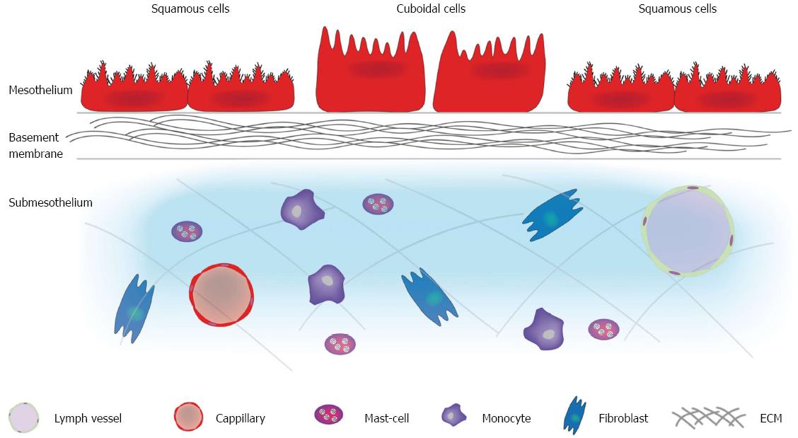
Figure 1 Structure of the peritoneum.
The peritoneum is composed of a mesothelium supported by a basement membrane that rests on a layer of submesothelium. The mesothelium consists of a monolayer of either flattened, stretch, squamous-like or cuboidal mesothelial cells. The luminal surface of mesothelial cells has numerous microvilli varying in shape, size and density. Cilia have also been identified on the surface of resting mesothelial cells. The basement membrane consists of a thin laminar network containing type I and IV collagen, proteoglycans and glycoproteins. The submesothelium consists of a complex network of extracellular matrix made up of different types of collagen, glycoproteins, glycosaminoglycans and proteoglycans. Blood vessels, lymphatics, and various cells types (fibroblasts, resident tissue macrophages, and mast cells) are also found is this layer. ECM: Extracellular matrix.
- Citation: Lemoine L, Sugarbaker P, Van der Speeten K. Pathophysiology of colorectal peritoneal carcinomatosis: Role of the peritoneum. World J Gastroenterol 2016; 22(34): 7692-7707
- URL: https://www.wjgnet.com/1007-9327/full/v22/i34/7692.htm
- DOI: https://dx.doi.org/10.3748/wjg.v22.i34.7692