Copyright
©The Author(s) 2016.
World J Gastroenterol. Sep 7, 2016; 22(33): 7507-7517
Published online Sep 7, 2016. doi: 10.3748/wjg.v22.i33.7507
Published online Sep 7, 2016. doi: 10.3748/wjg.v22.i33.7507
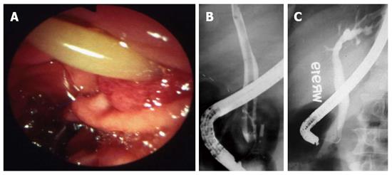
Figure 6 Duodenal ascariasis presenting as biliary colic.
A: Duodenoscopy showing adult ascaride in the ampullary orifice; B: Endoscopic retrograde cholangiogram showing long linear filling defect in the common bile duct; C: Cholangiogram after extraction of worms from bile duct. Patient had immediate relief of biliary colic. Adapted from Khuroo[5].
- Citation: Khuroo MS, Rather AA, Khuroo NS, Khuroo MS. Hepatobiliary and pancreatic ascariasis. World J Gastroenterol 2016; 22(33): 7507-7517
- URL: https://www.wjgnet.com/1007-9327/full/v22/i33/7507.htm
- DOI: https://dx.doi.org/10.3748/wjg.v22.i33.7507


