Copyright
©The Author(s) 2016.
World J Gastroenterol. Aug 28, 2016; 22(32): 7322-7331
Published online Aug 28, 2016. doi: 10.3748/wjg.v22.i32.7322
Published online Aug 28, 2016. doi: 10.3748/wjg.v22.i32.7322
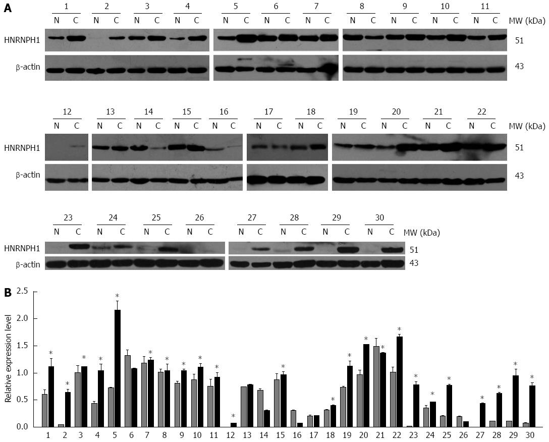
Figure 4 Western blotting analysis of heterogeneous nuclear ribonucleoprotein H1 in paired esophageal squamous cell carcinoma specimens.
A: Representative western blotting images of tumor (C) and matched adjacent non-tumor esophageal mucosal tissues (N) from 30 patients with ESCC. β-actin protein levels are shown as a loading control. The patients were coded from 1 to 30. B: Densitometric analysis of 30 ESCC cases. The gray and black bars represent the relative band intensity of HNRNPH1 in non-tumor (N) or tumor (C) tissues. Each data point represents the mean ± SD derived from three independent experiments. The asterisks mark the cases that overexpressed HNRNPH1 in tumor tissues. HNRNPH1: Heterogeneous nuclear ribonucleoprotein H1.
- Citation: Sun YL, Liu F, Liu F, Zhao XH. Protein and gene expression characteristics of heterogeneous nuclear ribonucleoprotein H1 in esophageal squamous cell carcinoma. World J Gastroenterol 2016; 22(32): 7322-7331
- URL: https://www.wjgnet.com/1007-9327/full/v22/i32/7322.htm
- DOI: https://dx.doi.org/10.3748/wjg.v22.i32.7322


