Copyright
©The Author(s) 2016.
World J Gastroenterol. Aug 21, 2016; 22(31): 7135-7145
Published online Aug 21, 2016. doi: 10.3748/wjg.v22.i31.7135
Published online Aug 21, 2016. doi: 10.3748/wjg.v22.i31.7135
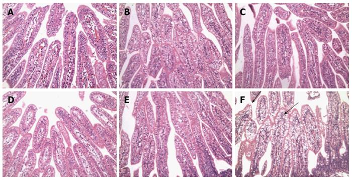
Figure 3 Histopathological lesions in the small intestine during rotavirus infection at 72 hpi.
Sections of the small intestine were stained with hematoxylin and eosin. Duodenal (A), jejunal (B), and ileal (C) mucosae collected from mock-infected control newborn mini-pigs or duodenal (D), jejunal (E), and ileal (F) mucosae that were collected from 5-d-old Group A virulent G1-impregnated newborn mini-pigs. Magnification, × 200.
- Citation: Li JT, Wei J, Guo HX, Han JB, Ye N, He HY, Yu TT, Wu YZ. Development of a human rotavirus induced diarrhea model in Chinese mini-pigs. World J Gastroenterol 2016; 22(31): 7135-7145
- URL: https://www.wjgnet.com/1007-9327/full/v22/i31/7135.htm
- DOI: https://dx.doi.org/10.3748/wjg.v22.i31.7135


