Copyright
©The Author(s) 2016.
World J Gastroenterol. Jan 21, 2016; 22(3): 917-932
Published online Jan 21, 2016. doi: 10.3748/wjg.v22.i3.917
Published online Jan 21, 2016. doi: 10.3748/wjg.v22.i3.917
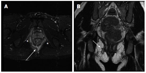
Figure 5 Perianal fistulizing Crohn’s disease depicted on magnetic resonance fistulography.
Axial short tau inversion recovery (A) and coronal (B) T2-weighted (B) images demonstrate a complex perianal fistula at the 6 o’clock position (A; arrow) with an intersphincteric component (A; asterisks) as well as a transphincteric track extending to the skin to the right of midline (B; arrow).
- Citation: Kilcoyne A, Kaplan JL, Gee MS. Inflammatory bowel disease imaging: Current practice and future directions. World J Gastroenterol 2016; 22(3): 917-932
- URL: https://www.wjgnet.com/1007-9327/full/v22/i3/917.htm
- DOI: https://dx.doi.org/10.3748/wjg.v22.i3.917


