Copyright
©The Author(s) 2016.
World J Gastroenterol. Jun 21, 2016; 22(23): 5422-5429
Published online Jun 21, 2016. doi: 10.3748/wjg.v22.i23.5422
Published online Jun 21, 2016. doi: 10.3748/wjg.v22.i23.5422
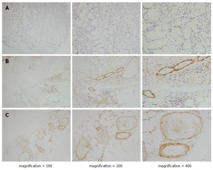
Figure 1 Immunohistochemical staining of endocan in gastric cancer.
Positive staining of endocan was observed in most of the endothelial cells of the tumour vessels, but less staining was observed in normal gastric tissues (A); In the peritumour vascular endothelium (B); positive endocan was more compact than that in the tumour centres (C).
- Citation: Chang Y, Niu W, Lian PL, Wang XQ, Meng ZX, Liu Y, Zhao R. Endocan-expressing microvessel density as a prognostic factor for survival in human gastric cancer. World J Gastroenterol 2016; 22(23): 5422-5429
- URL: https://www.wjgnet.com/1007-9327/full/v22/i23/5422.htm
- DOI: https://dx.doi.org/10.3748/wjg.v22.i23.5422


