Copyright
©The Author(s) 2016.
World J Gastroenterol. Jun 14, 2016; 22(22): 5154-5164
Published online Jun 14, 2016. doi: 10.3748/wjg.v22.i22.5154
Published online Jun 14, 2016. doi: 10.3748/wjg.v22.i22.5154
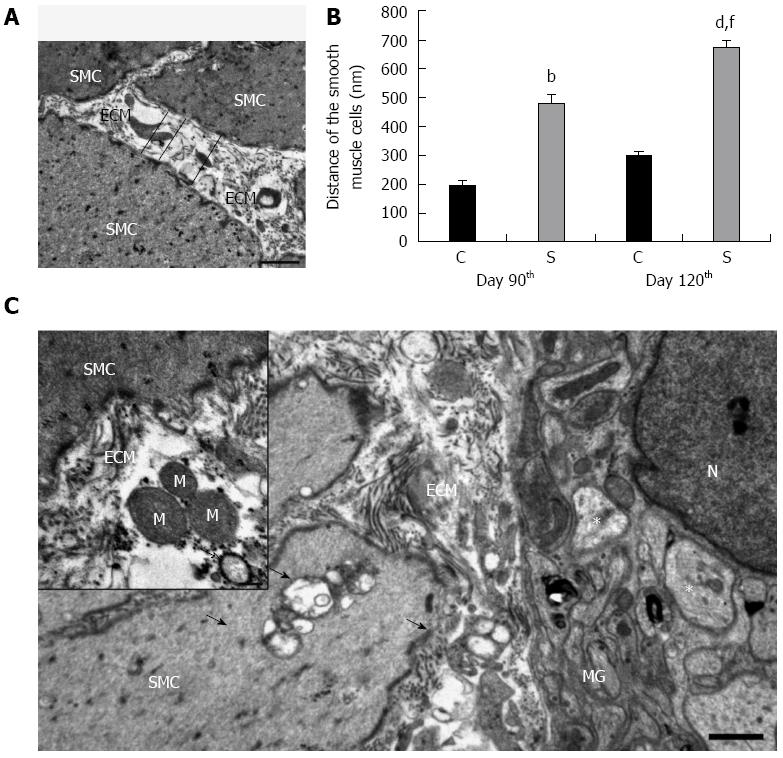
Figure 4 Ultrastructural alterations within the colon of control animals and in rats treated three times with 2,4,6-trinitrobenzenesulfonic acid.
Excess deposition of extracellular matrix (ECM) was observed within the smooth muscle layers in the ultrathin sections derived from the strictured region (A); Electronmicroscopic morphometry revealed that the distance between adjacent smooth muscle cells (SMCs) was significant larger in the strictured gut wall of the 2,4,6-trinitrobenzenesulfonic acid (TNBS)-treated rats (S) as compared with the gut wall of the control rats (C) on day 90 (B); A further significant increase in the mean separation distance of the SMCs was recorded on day 120 post-TNBS treatment (B). Data are expressed as mean ± SE. bP < 0.01, dP < 0.001 TNBS-treated groups vs age-matched controls; fP < 0.001 TNBS-treated group on the day 90 vs TNBS-treated group on day 120. Representative electron micrograph of the strictured colonic area 120 d after the third TNBS administration (C). Because of ECM accumulation, the SMCs also moved away from the myenteric ganglia (MGs). Swollen and empty confluent vacuoles of different sizes (arrows) were frequently seen in the SMCs and also in their close environment. Rupture of the plasma membrane and subsequent leakage of the cell organelles into the microenvironment, e.g., the mitochondria (M) and autophagosomes (hollow arrow), were frequently seen in the intercellular spaces (insert). However, the vast majority of the axons appeared normal; necrotic axons were rarely seen in the MGs (asterisks). N: Nucleus. Bars: 1 μm and 200 nm (insert).
- Citation: Talapka P, Berkó A, Nagy LI, Chandrakumar L, Bagyánszki M, Puskás LG, Fekete &, Bódi N. Structural and molecular features of intestinal strictures in rats with Crohn's-like disease. World J Gastroenterol 2016; 22(22): 5154-5164
- URL: https://www.wjgnet.com/1007-9327/full/v22/i22/5154.htm
- DOI: https://dx.doi.org/10.3748/wjg.v22.i22.5154


