Copyright
©The Author(s) 2016.
World J Gastroenterol. May 28, 2016; 22(20): 4901-4907
Published online May 28, 2016. doi: 10.3748/wjg.v22.i20.4901
Published online May 28, 2016. doi: 10.3748/wjg.v22.i20.4901
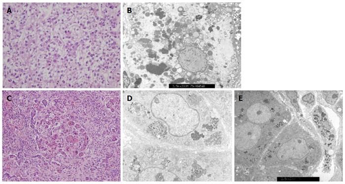
Figure 2 Liver histologic features from infants with chronic intrahepatic cholestasis with normal ranges of γ-glutamyl transpeptidase.
A: Hepatocellular carcinoma was confirmed by liver specimen at hepatectomy taken from patient 1 at 24 mo of age. Cellular atypia with trabecular and acinar type was shown. Microvascular invasion was not identified; hematoxylin-eosin stain, original magnification × 400; B: Electron microscopic examination of liver specimen from patient 2 shows many globular or curly appearance electron dense materials in the cytoplasm with original magnification of × 3.5k; C: Liver biopsy at hepatectomy taken at 6 months of age from patient 3 shows periportal fibrosis, inflammatory cell infiltration, intracanalicular bile plugs, giant cell formation, and bile ductular proliferation; hematoxylin-eosin stain, original magnification × 200; D: Electron microscopic examination of liver specimen from patient 4 at 3.5 mo of age reveals amorphous and coarse granular bile pigments in the dilated bile canaliculi. Original magnification × 5.0k; E: Electron microscopic examination of liver specimen from patient 5 shows aggregated bile pigments in the cytoplasm. Original magnification × 2.5k.
- Citation: Park JS, Ko JS, Seo JK, Moon JS, Park SS. Clinical and ABCB11 profiles in Korean infants with progressive familial intrahepatic cholestasis. World J Gastroenterol 2016; 22(20): 4901-4907
- URL: https://www.wjgnet.com/1007-9327/full/v22/i20/4901.htm
- DOI: https://dx.doi.org/10.3748/wjg.v22.i20.4901


