Copyright
©The Author(s) 2016.
World J Gastroenterol. May 28, 2016; 22(20): 4881-4890
Published online May 28, 2016. doi: 10.3748/wjg.v22.i20.4881
Published online May 28, 2016. doi: 10.3748/wjg.v22.i20.4881
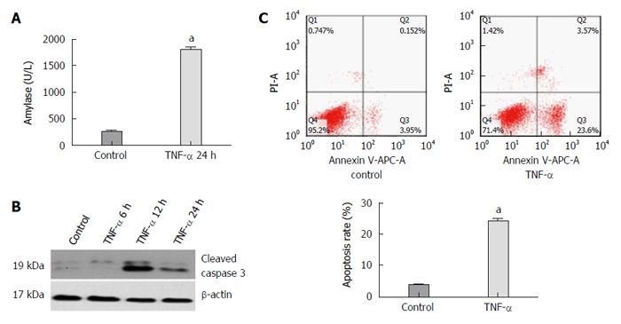
Figure 3 Expression of amylase, activated caspase 3 protein, apoptosis rate of AR42J cells and miR-29a level increase in the experimental group compared with the control group.
A: The expression of amylase analysis in the supernatant; B: Western blot analysis of activated caspase 3 in AR42J cells; C: The apoptosis rate of AR42J cells after the treatment with TNF-α for 24 h. Date were obtained from three independent experiments in triplicate and are shown as the mean ± SD. aP < 0.05 vs control group.
- Citation: Fu Q, Qin T, Chen L, Liu CJ, Zhang X, Wang YZ, Hu MX, Chu HY, Zhang HW. miR-29a up-regulation in AR42J cells contributes to apoptosis via targeting TNFRSF1A gene. World J Gastroenterol 2016; 22(20): 4881-4890
- URL: https://www.wjgnet.com/1007-9327/full/v22/i20/4881.htm
- DOI: https://dx.doi.org/10.3748/wjg.v22.i20.4881


