Copyright
©The Author(s) 2016.
World J Gastroenterol. May 14, 2016; 22(18): 4515-4528
Published online May 14, 2016. doi: 10.3748/wjg.v22.i18.4515
Published online May 14, 2016. doi: 10.3748/wjg.v22.i18.4515
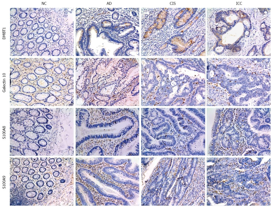
Figure 5 Representative results of immunohistochemistry show the expression of DMBT1, S100A9, Galectin-10 and S100A8 in the NC, AD, CIS and ICC (Original magnification, × 200).
DMBT1 immunostaining in NC, AD, CIS and ICC. Negative staining was observed in NC, moderate in AD tissues, and strong cytoplasmic staining in CIS and ICC tissues. S100A9 immunostaining in NC, AD, CIS and ICC. Negative staining was found in NC, weak intralesional staining in AD, moderate intralesional staining in CIS and strong intralesional staining in ICC. Galectin-10 immunostaining in NC, AD, CIS and ICC. Negative staining was found in NC, weak staining in AD and CIS, and moderate staining in ICC. S100A8 immunostaining in NC, AD, CIS and ICC. Negative staining was observed in NC, moderate intralesional staining in AD and CIS, and strong intralesional staining in ICC.
- Citation: Peng F, Huang Y, Li MY, Li GQ, Huang HC, Guan R, Chen ZC, Liang SP, Chen YH. Dissecting characteristics and dynamics of differentially expressed proteins during multistage carcinogenesis of human colorectal cancer. World J Gastroenterol 2016; 22(18): 4515-4528
- URL: https://www.wjgnet.com/1007-9327/full/v22/i18/4515.htm
- DOI: https://dx.doi.org/10.3748/wjg.v22.i18.4515


