Copyright
©The Author(s) 2016.
World J Gastroenterol. May 14, 2016; 22(18): 4466-4483
Published online May 14, 2016. doi: 10.3748/wjg.v22.i18.4466
Published online May 14, 2016. doi: 10.3748/wjg.v22.i18.4466
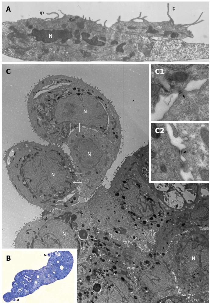
Figure 4 PL45 ultrastructure.
A: Electron microscopy of two PL45 cells grown in 2D-monolayers that exhibit protrusions consistent with invadopodia (ip) originating from their surface and projecting toward the culture medium. N: Nucleus. Scale bar = 1 μm; B: Light microscopy of a semi-thin section showing a multilayered spheroid with 2 small groups of cells partially detached from the periphery of the spheroid (arrows). Scale bar = 40 μm; C: Electron microscopy of a thin section adjacent to the semi-thin one showing a group consisting of 3 cells joined to each other. The outlined areas are shown at greater enlargement in inserts C1 and C2; arrows indicate desmosomes. N: Nucleus. Scale bar = 5 μm. Inset scale bar = 0.25 μm.
- Citation: Gagliano N, Celesti G, Tacchini L, Pluchino S, Sforza C, Rasile M, Valerio V, Laghi L, Conte V, Procacci P. Epithelial-to-mesenchymal transition in pancreatic ductal adenocarcinoma: Characterization in a 3D-cell culture model. World J Gastroenterol 2016; 22(18): 4466-4483
- URL: https://www.wjgnet.com/1007-9327/full/v22/i18/4466.htm
- DOI: https://dx.doi.org/10.3748/wjg.v22.i18.4466


