Copyright
©The Author(s) 2016.
World J Gastroenterol. May 7, 2016; 22(17): 4411-4415
Published online May 7, 2016. doi: 10.3748/wjg.v22.i17.4411
Published online May 7, 2016. doi: 10.3748/wjg.v22.i17.4411
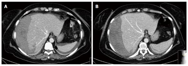
Figure 1 Urgent abdomen computed tomography scan.
A: Hepatic subcapsular hematoma of the right lobe (14 cm × 6 cm × 19 cm) with peripheral parenchymal laceration. Ab-extrinsic compression of the right and middle hepatic vein with perisplenic free fluid; B: Six days after radiological selective embolization: note the stability of the haematoma dimension with disappearance of perisplenic free fluid.
- Citation: Zappa MA, Aiolfi A, Antonini I, Musolino CD, Porta A. Subcapsular hepatic haematoma of the right lobe following endoscopic retrograde cholangiopancreatography: Case report and literature review. World J Gastroenterol 2016; 22(17): 4411-4415
- URL: https://www.wjgnet.com/1007-9327/full/v22/i17/4411.htm
- DOI: https://dx.doi.org/10.3748/wjg.v22.i17.4411


