Copyright
©The Author(s) 2016.
World J Gastroenterol. Apr 28, 2016; 22(16): 4149-4159
Published online Apr 28, 2016. doi: 10.3748/wjg.v22.i16.4149
Published online Apr 28, 2016. doi: 10.3748/wjg.v22.i16.4149
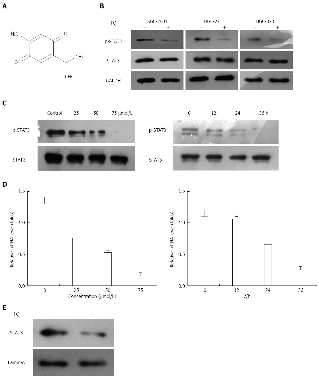
Figure 1 Thymoquinone suppresses constitutive STAT3 activation in three gastric cancer cell lines.
A: Human gastric cancer cells (HGC27, BGC823, and SGC7901) were incubated with 50 μmol/L thymoquinone (TQ) for 24 h, and western blotting was performed as described previously; B: The same blots were stripped and reprobed with the GAPDH antibody to verify equal protein loading; C: TQ suppressed phospho-STAT3 levels in a concentration- and time-dependent manner. HGC27 cells (1 × 10⁶/mL) were treated with the indicated concentrations of TQ for 24 h, and western blotting was performed (left). HGC27 cells (1 × 10⁶/mL) were treated with 50 μmol/L TQ for the indicated times, and western blotting was performed (right). The same blots were stripped and reprobed with the STAT3 antibody to verify equal protein loading; D: TQ suppressed STAT3 gene levels in a concentration- and time-dependent manner. HGC27 cells (1 × 10⁶/mL) were treated with the indicated concentrations of TQ for 24 h, and real-time RT-PCR was performed (left). HGC27 cells (1 × 10⁶/mL) were treated with 50 μmol/L TQ for the indicated times, and real-time RT-PCR was performed (right); E: TQ suppressed STAT3 nuclear translocation. HGC27 cells (1 × 10⁶/mL) cells were treated with or without 50 μmol/L TQ for 24h, and nuclear extracts were assessed for the detection nuclear accumulation of STAT3. Lamin-A was used as a nuclear extract marker. GAPDH: Glyceraldehyde-3-phosphate dehydrogenase; RT-PCR: Reverse transcription polymerase chain reaction; STAT: Signal transducer and activator of transcription.
- Citation: Zhu WQ, Wang J, Guo XF, Liu Z, Dong WG. Thymoquinone inhibits proliferation in gastric cancer via the STAT3 pathway in vivo and in vitro. World J Gastroenterol 2016; 22(16): 4149-4159
- URL: https://www.wjgnet.com/1007-9327/full/v22/i16/4149.htm
- DOI: https://dx.doi.org/10.3748/wjg.v22.i16.4149


