Copyright
©The Author(s) 2016.
World J Gastroenterol. Apr 21, 2016; 22(15): 4027-4033
Published online Apr 21, 2016. doi: 10.3748/wjg.v22.i15.4027
Published online Apr 21, 2016. doi: 10.3748/wjg.v22.i15.4027
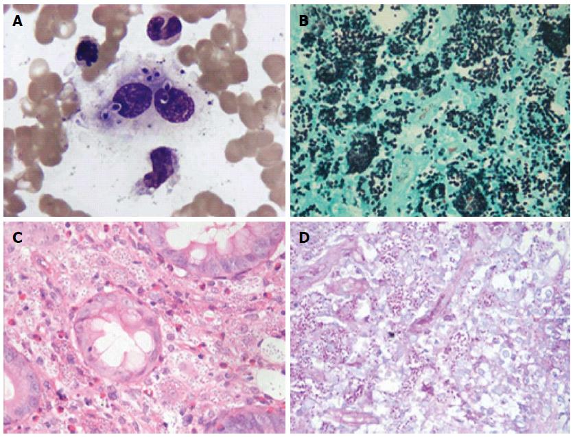
Figure 1 Pathological stain results of Histoplasma capsulatum in bone marrow.
A: Wright-Giemsa stained bone marrow aspirate. There were numerous round or oval H. capsulatum of relatively uniform size in phagocyte and cytoplasm. It is round at one end and pointed at the other. Karyon was stained fuchsia, surrounded by peri-nuclear halos and the shape was capsule-like; B-D: Numerous uniform oval-shaped yeasts suggesting H. capsulatum were found in the amina propria stroma in the descending colon biopsy. [B: Gomori methenamine silver stain (magnification × 100); C: Hematoxylin and eosin stain (magnification × 40); D: Periodic acid-Schiff stain (magnification × 40)]. H. capsulatum: Histoplasma capsulatum.
- Citation: Zhu LL, Wang J, Wang ZJ, Wang YP, Yang JL. Intestinal histoplasmosis in immunocompetent adults. World J Gastroenterol 2016; 22(15): 4027-4033
- URL: https://www.wjgnet.com/1007-9327/full/v22/i15/4027.htm
- DOI: https://dx.doi.org/10.3748/wjg.v22.i15.4027


