Copyright
©The Author(s) 2016.
World J Gastroenterol. Apr 14, 2016; 22(14): 3735-3745
Published online Apr 14, 2016. doi: 10.3748/wjg.v22.i14.3735
Published online Apr 14, 2016. doi: 10.3748/wjg.v22.i14.3735
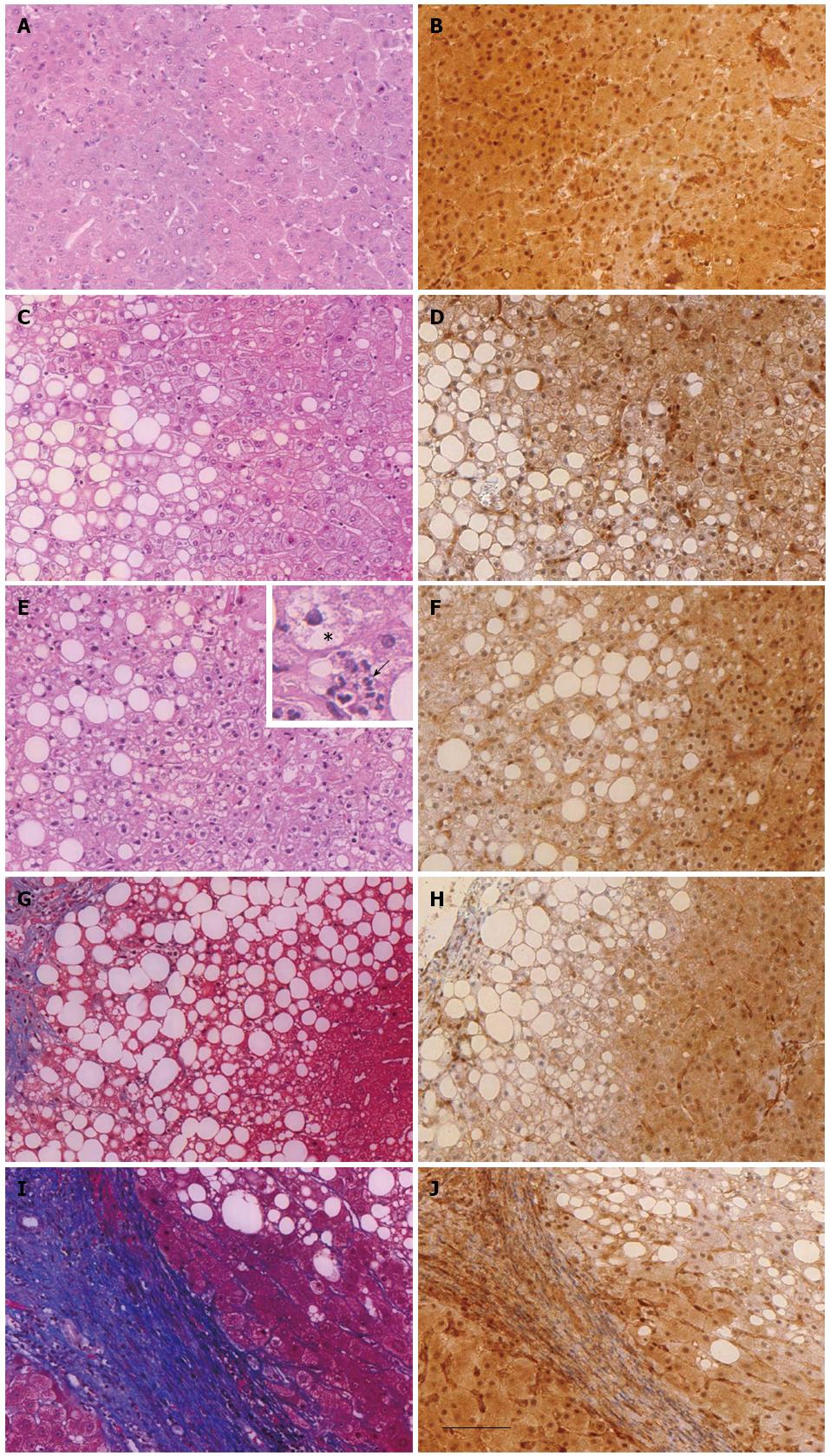
Figure 1 General histology and immunohistochemical detection of phosphatase and tensin homolog protein expression in the liver of healthy donor (hepatic resections) or patients with different stages of non-alcoholic fatty liver disease (liver biopsies).
Liver sections of healthy donors (A and B) or of obese patients with steatosis (n = 10, C and D), nonalcoholic steatohepatitis (n = 14, E and F), fibrosis (n = 12, G and H) and cirrhosis (n = 8, I and J) were either stained with hematoxylin eosin (A, C, E), Masson’s trichrome (G and I) or immunostained with anti-PTEN antibody (B, D, F, H and J). The inset in image (E) shows hepatic intralobular inflammation characterized by neutrophils (arrow) and ballooning hepatocyte (star). Scale bar = 100 μm. PTEN: Phosphatase and tensin homolog.
- Citation: Sanchez-Pareja A, Clément S, Peyrou M, Spahr L, Negro F, Rubbia-Brandt L, Foti M. Phosphatase and tensin homolog is a differential diagnostic marker between nonalcoholic and alcoholic fatty liver disease. World J Gastroenterol 2016; 22(14): 3735-3745
- URL: https://www.wjgnet.com/1007-9327/full/v22/i14/3735.htm
- DOI: https://dx.doi.org/10.3748/wjg.v22.i14.3735


