Copyright
©The Author(s) 2016.
World J Gastroenterol. Apr 7, 2016; 22(13): 3687-3692
Published online Apr 7, 2016. doi: 10.3748/wjg.v22.i13.3687
Published online Apr 7, 2016. doi: 10.3748/wjg.v22.i13.3687
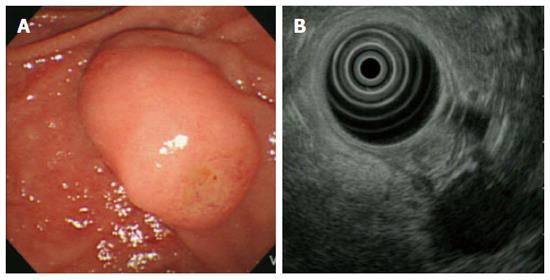
Figure 1 A duodenoscopic image showed an enlarged major papilla with central umbilication and fine nodularity on ampullary orifice (A), endoscopic ultrasonography at the major ampulla revealed a 1.
1 × 0.9-cm, slightly hypoechoic round ampullary mass confined to the submucosa without definite wall disruption or adjacent invasion (B).
- Citation: Lee SH, Lee TH, Jang SH, Choi CY, Lee WM, Min JH, Cho HD, Park SH. Ampullary neuroendocrine tumor diagnosed by endoscopic papillectomy in previously confirmed ampullary adenoma. World J Gastroenterol 2016; 22(13): 3687-3692
- URL: https://www.wjgnet.com/1007-9327/full/v22/i13/3687.htm
- DOI: https://dx.doi.org/10.3748/wjg.v22.i13.3687


