Copyright
©The Author(s) 2016.
World J Gastroenterol. Jan 7, 2016; 22(1): 89-102
Published online Jan 7, 2016. doi: 10.3748/wjg.v22.i1.89
Published online Jan 7, 2016. doi: 10.3748/wjg.v22.i1.89
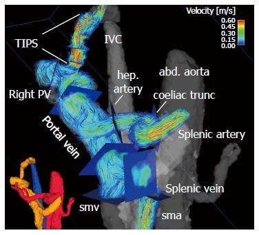
Figure 3 Four-dimensional flow magnetic resonance imaging in a 63-yr-old female with liver cirrhosis after TIPS placement.
Small lower left corner: three-dimensional (3D) segmentation of the liver and splanchnic hemodynamics (red: arterial system; orange: portal venous system; blue: venous system). Large figure: Color-coded 3D particle-traces visualization demonstrates increased velocities within the TIPS and the arteries. Blue analysis planes were positioned throughout the arterial and portal venous systems as well as TIPS to quantify liver hemodynamics. abd. aorta: Abdominal aorta; hep. artery: Hepatic artery; sma: Superior mesenteric artery; smv: Superior mesenteric vein; Right PV: Right portal vein branch; IVC: Inferior vena cava.
- Citation: Stankovic Z. Four-dimensional flow magnetic resonance imaging in cirrhosis. World J Gastroenterol 2016; 22(1): 89-102
- URL: https://www.wjgnet.com/1007-9327/full/v22/i1/89.htm
- DOI: https://dx.doi.org/10.3748/wjg.v22.i1.89


