Copyright
©The Author(s) 2016.
World J Gastroenterol. Jan 7, 2016; 22(1): 284-299
Published online Jan 7, 2016. doi: 10.3748/wjg.v22.i1.284
Published online Jan 7, 2016. doi: 10.3748/wjg.v22.i1.284
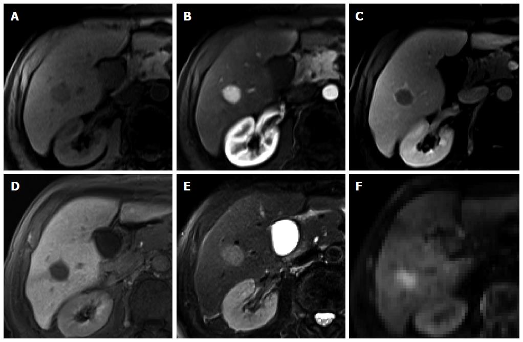
Figure 1 Hepatocellular carcinoma in a 74-year-old man with hepatitis C infection.
A: Precontrast T1-weighted image shows a hypointense nodule in segment 6; B: Hepatic arterial phase of gadoxetic acid-enhanced MRI shows homogeneous marked enhancement of the tumor; C: Transitional phase shows washout of the contrast medium in the tumor with capsular enhancement; D: Hepatobiliary phase shows marked hypointensity of the tumor relative to the liver parenchyma; E, F: T2-weighted image and diffusion weighted image (b = 800) show high SI of the tumor.
- Citation: Park YS, Lee CH, Kim JW, Shin S, Park CM. Differentiation of hepatocellular carcinoma from its various mimickers in liver magnetic resonance imaging: What are the tips when using hepatocyte-specific agents? World J Gastroenterol 2016; 22(1): 284-299
- URL: https://www.wjgnet.com/1007-9327/full/v22/i1/284.htm
- DOI: https://dx.doi.org/10.3748/wjg.v22.i1.284


