Copyright
©The Author(s) 2015.
World J Gastroenterol. Mar 7, 2015; 21(9): 2820-2825
Published online Mar 7, 2015. doi: 10.3748/wjg.v21.i9.2820
Published online Mar 7, 2015. doi: 10.3748/wjg.v21.i9.2820
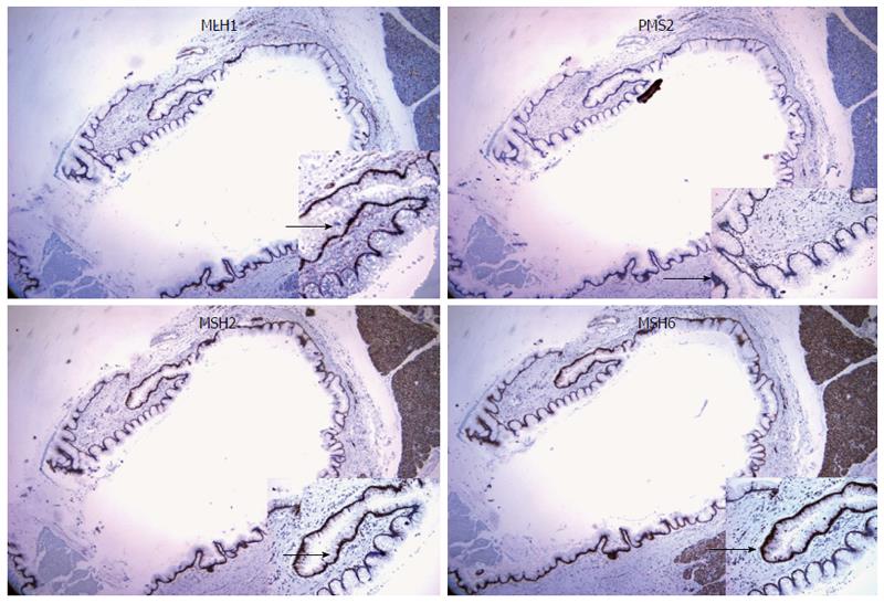
Figure 4 Immunohistochemical staining.
Staining for MLH1, PMS2, MSH2, and MSH6 did not reveal loss of mismatch repair protein expression, as the neoplastic cells show uniform nuclear stain (black arrows) for all four markers (40 × magnification).
- Citation: Flanagan MR, Jayaraj A, Xiong W, Yeh MM, Raskind WH, Pillarisetty VG. Pancreatic intraductal papillary mucinous neoplasm in a patient with Lynch syndrome. World J Gastroenterol 2015; 21(9): 2820-2825
- URL: https://www.wjgnet.com/1007-9327/full/v21/i9/2820.htm
- DOI: https://dx.doi.org/10.3748/wjg.v21.i9.2820


