Copyright
©The Author(s) 2015.
World J Gastroenterol. Mar 7, 2015; 21(9): 2820-2825
Published online Mar 7, 2015. doi: 10.3748/wjg.v21.i9.2820
Published online Mar 7, 2015. doi: 10.3748/wjg.v21.i9.2820
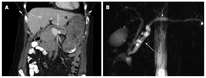
Figure 1 Pancreatic imaging.
A: Computed tomography imaging demonstrated progressive dilation of main pancreatic duct as compared to the prior year (white arrow). B: Magnetic resonance cholangiopancreatography shows pancreatic ductal dilation with a normal biliary tree (short white arrow). There is evidence of side-branch dilation within the head and uncinate process (long white arrow).
- Citation: Flanagan MR, Jayaraj A, Xiong W, Yeh MM, Raskind WH, Pillarisetty VG. Pancreatic intraductal papillary mucinous neoplasm in a patient with Lynch syndrome. World J Gastroenterol 2015; 21(9): 2820-2825
- URL: https://www.wjgnet.com/1007-9327/full/v21/i9/2820.htm
- DOI: https://dx.doi.org/10.3748/wjg.v21.i9.2820


