Copyright
©The Author(s) 2015.
World J Gastroenterol. Feb 14, 2015; 21(6): 1915-1926
Published online Feb 14, 2015. doi: 10.3748/wjg.v21.i6.1915
Published online Feb 14, 2015. doi: 10.3748/wjg.v21.i6.1915
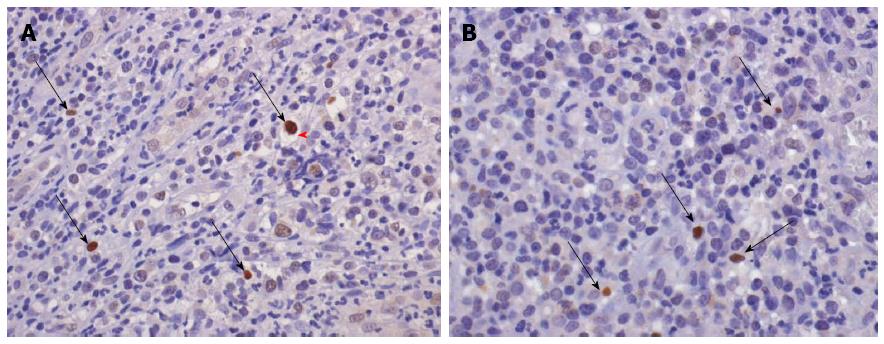
Figure 3 Pathognomonic “cytomegalic” cell (i.
e., enlarged cell surrounded by a light-coloured halo, red arrow) with a brown reactive nuclear inclusion (thin black arrows) and a few scattered positive cells for the human Cytomegalovirus are shown (panel A, immunoperoxidase-hematoxylin, original magnification × 250). Some positive cells with brown nuclei (thin black arrows) are evident following the specific staining for the Epstein-Barr virus nuclear antigen-1 (panel B, immunoperoxidase-hematoxylin, original magnification × 400).
- Citation: Ciccocioppo R, Racca F, Paolucci S, Campanini G, Pozzi L, Betti E, Riboni R, Vanoli A, Baldanti F, Corazza GR. Human cytomegalovirus and Epstein-Barr virus infection in inflammatory bowel disease: Need for mucosal viral load measurement. World J Gastroenterol 2015; 21(6): 1915-1926
- URL: https://www.wjgnet.com/1007-9327/full/v21/i6/1915.htm
- DOI: https://dx.doi.org/10.3748/wjg.v21.i6.1915


