Copyright
©The Author(s) 2015.
World J Gastroenterol. Feb 7, 2015; 21(5): 1468-1478
Published online Feb 7, 2015. doi: 10.3748/wjg.v21.i5.1468
Published online Feb 7, 2015. doi: 10.3748/wjg.v21.i5.1468
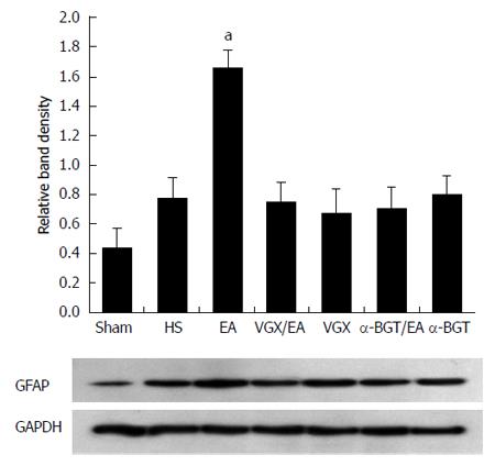
Figure 2 Glial fibrillary acidic protein expression at 6 h after blood loss.
Intestinal extracts were obtained from rats at 6 h after blood loss for measurement of glial fibrillary acidic protein expression using western blotting. Representative blots for glial fibrillary acidic protein (GFAP) are shown, with the corresponding GAPDH loading control to demonstrate equal protein load in all lanes. The GFAP level was lower in the sham group. EA ST36 increased GFAP expression (aP < 0.05 vs HS group). Significantly weak GFAP expression was seen in all the other injury groups (n = 3-6 rats per group at 6 h after blood loss). α-BGT: α-bungarotoxin; VGX: Vagotomy; EA: Electroacupuncture; HS: Hemorrhagic shock.
- Citation: Hu S, Zhao ZK, Liu R, Wang HB, Gu CY, Luo HM, Wang H, Du MH, Lv Y, Shi X. Electroacupuncture activates enteric glial cells and protects the gut barrier in hemorrhaged rats. World J Gastroenterol 2015; 21(5): 1468-1478
- URL: https://www.wjgnet.com/1007-9327/full/v21/i5/1468.htm
- DOI: https://dx.doi.org/10.3748/wjg.v21.i5.1468


