Copyright
©The Author(s) 2015.
World J Gastroenterol. Feb 7, 2015; 21(5): 1468-1478
Published online Feb 7, 2015. doi: 10.3748/wjg.v21.i5.1468
Published online Feb 7, 2015. doi: 10.3748/wjg.v21.i5.1468
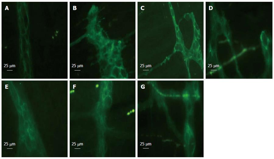
Figure 1 Glial fibrillary acidic protein immunofluorescent staining at 6 h after blood loss.
Enteric glial cells (EGCs) were distorted following hemorrhage and showed morphological abnormalities. EGCs presented as normal in the sham group. Electroacupuncture (EA) ST36 attenuated the morphological changes. After hemorrhagic shock (HS), the fluorescent intensity was enhanced. EA ST36 showed more intense fluorescence of glial fibrillary acidic protein (GFAP) staining, whereas fluorescence after VGX or injection of α-bungarotoxin (BGT) was weak. All images were taken at magnification × 400; white bar = 25 μm (n = 3-6 rats per group at 6 h after blood loss). A: Sham group; B: HS group; C: EA group; D: VGX/EA group; E: VGX group; F: α-BGT/EA; G: α-BGT group.
- Citation: Hu S, Zhao ZK, Liu R, Wang HB, Gu CY, Luo HM, Wang H, Du MH, Lv Y, Shi X. Electroacupuncture activates enteric glial cells and protects the gut barrier in hemorrhaged rats. World J Gastroenterol 2015; 21(5): 1468-1478
- URL: https://www.wjgnet.com/1007-9327/full/v21/i5/1468.htm
- DOI: https://dx.doi.org/10.3748/wjg.v21.i5.1468


