Copyright
©The Author(s) 2015.
World J Gastroenterol. Dec 14, 2015; 21(46): 13201-13204
Published online Dec 14, 2015. doi: 10.3748/wjg.v21.i46.13201
Published online Dec 14, 2015. doi: 10.3748/wjg.v21.i46.13201
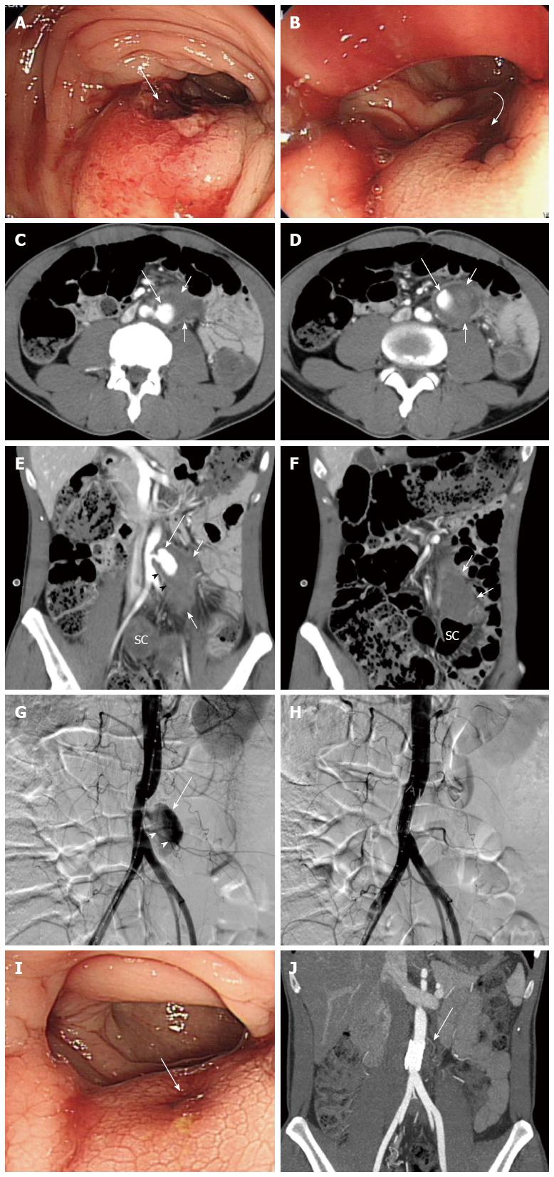
Figure 1 Thirty seven-year-old male with Behçet’s disease.
A, B: Sigmoidoscopy revealed a submucosal tumor-like protrusion (long arrow in A) with a bleeding ulceration (curved arrow in B) in the proximal sigmoid colon; C-F: Axial (C, D) and coronal reformatted (E, F) images of a contrast-enhanced computed tomography (CT) scan revealing an enhanced pseudoaneurysm (long arrows in C-E) above the origin site of the inferior mesenteric artery (arrowheads in E) with a hematoma (short arrows in C-F) extending into the subperitoneal space of the sigmoid mesocolon. Hyperdense clotted blood was also noted in the lumen of the sigmoid colon (SC); G: Frontal abdominal aortogram revealing a pseudoaneurysm (arrow) above the origin site of the inferior mesenteric artery (arrowheads); H: Abdominal aortogram following stent grafting indicating disappearance of the pseudoaneurysm; I: Follow-up sigmoidoscopy (4 d after the procedure) shows mild mucosal elevation with erosion (arrow) and no evidence of active bleeding; J: Follow-up computed tomography angiography (6 mo after the procedure) demonstrated the aortic stent grafting state (arrow) and complete remission of the aortic pseudoaneurysm and hematoma in the sigmoid mesocolon without evidence of graft infection.
- Citation: Lee SL, Ku YM, Won Y. Spontaneous aortic pseudoaneurysm rupture into the sigmoid colon in Behçet’s disease patient. World J Gastroenterol 2015; 21(46): 13201-13204
- URL: https://www.wjgnet.com/1007-9327/full/v21/i46/13201.htm
- DOI: https://dx.doi.org/10.3748/wjg.v21.i46.13201


